Speaker
Description
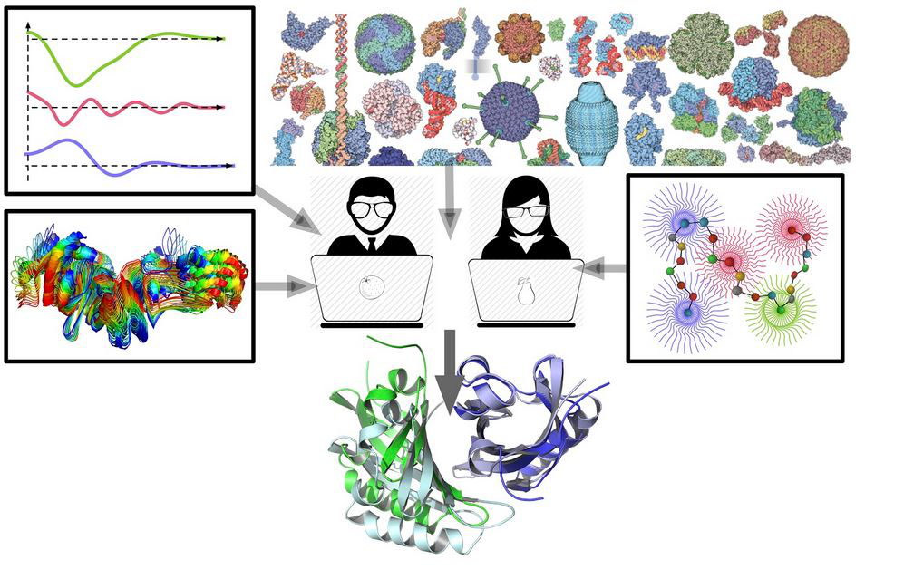
While crystallography has been providing atomic-resolution structures of biomolecules for over half a century, the real challenge of today’s biophysics is to correlate molecules’ structure and dynamics in solution with their function. Owing to the complexity of the problem, the answer can only be found if multiple sources of information are used simultaneously. I will present a set of tools developed for this purpose in our group [1].
Recently, we introduced SAXS and SANS packages called Pepsi-SAXS, and Pepsi-SANS, correspondingly [1,2]. Pepsi-SAXS is a very efficient method that calculates small angle X-ray scattering profiles from atomistic models. It is based on the multipole expansion scheme and is significantly faster than other methods with the same level of precision. One of the challenges in the field is, however, flexible fitting of atomistic models into small-angle scattering profiles. We designed a computational scheme that uses the nonlinear normal modes [1,3] as a low-dimensional representation of the protein motion subspace and optimizes protein structures guided by the SAXS and SANS profiles. For example, this scheme was ranked first in the recent CASP13 structure prediction challenge. Another challenge is data-assisted protein docking. We have designed a scheme for SAXS-assisted rescoring of docking predictions. This was made possible due to the polynomial representation of partial scattering amplitudes for each of the docking partners.
We are also interested in protein symmetry, as many protein complexes are symmetric homooligomers. We have designed a novel free-docking symmetry-assisted method [4] and also a dual analytical technique for identification of symmetry in a protein assembly [5-7]. This allowed us to study principles of protein organizations on the PDB scale.
Finally, artificial intelligence has made a big leap forward and found many applications in structural bioinformatics. On our side, we have been using it for multiple tasks of protein structure prediction, including protein-protein [8] and protein-ligand [9] docking, shape analysis [7] and structure prediction [10-11].
References
[1] - https://team.inria.fr/nano-d/software/.
[2] - S. Grudinin, et al, Acta Cryst D, (2017), D3, 449.
[3] - A. Hoffmann & S. Grudinin, J. Chem .Theory Comput., 2017, 13 (5), 2123.
[4] - Ritchie, D. W. & Grudinin, S (2016). J. Appl. Cryst., 49, 1-10.
[5] - Pages, G., Kinzina, E, & Grudinin, S (2018). J. Struct.Biology, 203 (2), 142-148.
[6] - Pages, G. & Grudinin, S (2018). J. Struct.Biology, 203 (3), 185-194.
[7] - Pages, G. & Grudinin, S (2018). arXiv preprint arXiv:1810.12026.
[8] - Neveu, E., D. Ritchie, P. Popov, & S. Grudinin (2016). Bioinformatics, 32 (7), pp.i693-i701.
[9] - Kadukova, M. & S. Grudinin (2017). J. Comp.-Aid. Mol. Des. 31 (10), pp.943-958.
[10] - Karasikov, M., G. Pagès, & S.Grudinin (2018), Bioinformatics, bty1037.
[11] - Pages, G., B. Charmettant & S. Grudinin (2019). Bioinformatics, btz122.

