canSAS 2023

---------------------------------------------------
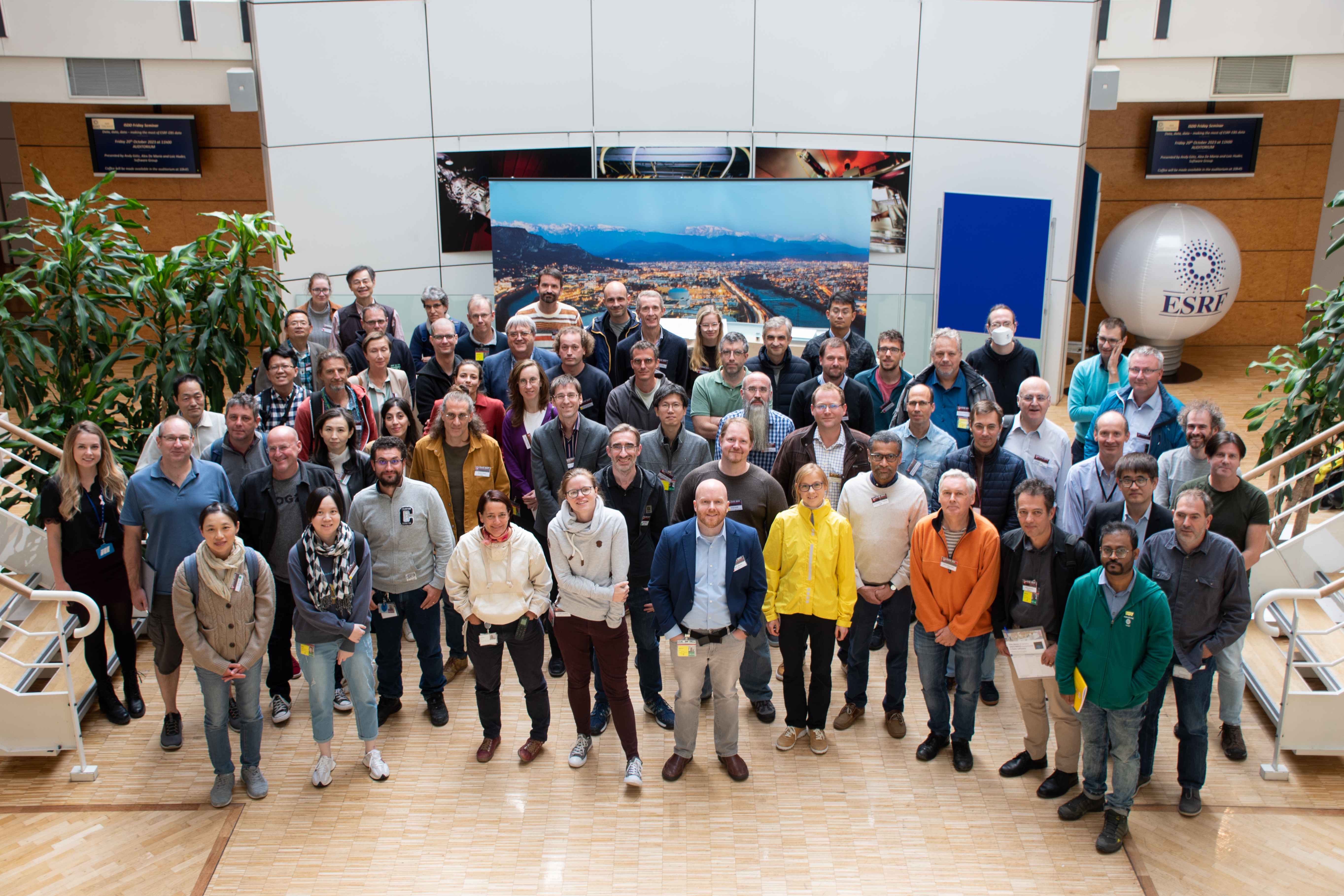
The 25th anniversary workshop of canSAS will be held in Grenoble from Monday 16th - Wednesday 18th October 2023.
The acronym canSAS stands for collective action for nomadic Small Angle Scatterers. It is a bottom-up effort originally initiated in Grenoble by Ron Ghosh (ILL), Roland May (ILL), Claudio Ferrero (ESRF) and Wim Bras (DUBBLE CRG) in 1998. Ever since it has been an ongoing activity to provide the small-angle scattering user community with shared tools for data reduction and analysis, and disseminating other pertinent information (https://www.cansas.org).
Lately the focus has been on data analysis and modeling software, instrument resolution corrections and other issues related to data interpretation.
The last formal workshop was held in Freising, Germany in 2019
(https://www.cansas.org/wgwiki/index.php/canSAS-XI). The following meeting planned in Delaware could not be organized due to the pandemic. This workshop will be useful in particular for instrument scientists and users who are deeply involved in development of new software tools or methods.
https://wiki.cansas.org/index.php/canSAS
The following topics are expected to be covered during the workshop:
- ML based SAS data analysis
- Emerging sample environments and methods
- SAS based imaging (tensor tomography, USAS imaging, ptychography, CXDI, etc.)
- Simultaneous SANS/SAXS fits, AI based methods for data collection and data mining, Refraction/Reflection effects)
- Needs and developments of analysis programs
- Background corrections, anisotropic scattering, fiber diffraction)
- Open access, open data, enhancing collaboration between facilities worldwide
We would like to extend our thanks to sas2024 for supporting our event.
- AdrianRennie_CanSAS2023.pdf
- AnneMartel_CanSAS2023.pptx
- ClementBlanchet_CanSAS2023.pdf
- CyJeffries_CanSAS2023.pdf
- FrancescoSpinozzi_CanSAS2023.pdf
- HenrichFrielinghaus_CanSAS2023.pptx
- OrionShih_CanSAS2023.pdf
- ShunYu_CanSAS2023.pdf
- StephenKing_CanSAS2023.pdf
- TimSnow_CanSAS2023.pdf
- UriRaviv_CanSAS2023.pdf
- WeiRenChen_CanSAS2023.pptx
- WiebkeKoepp_CanSAS2023.pdf
- WojciechPotrzebowski_CanSAS2023.pdf
- YoshiharuNishiyama_CanSAS2023.pdf
-
-
11:30
→
12:00
Registration
-
12:00
→
13:30
Lunch 1h 30m
-
13:30
→
14:00
Opening ILL/ESRF
-
13:30
Introductory words by ILL Science Director Jacques Jestin 15m
-
13:45
Introductory words by T. Narayanan on behalf of ESRF Director of Research (ad interim) Michael Krisch 15m
-
13:30
-
14:00
→
16:00
Machine learning based SAS data analysis: Chair: Sylvain Prévost + Narayanan Theyencheri
-
14:00
The Autonomous Formulation Lab: Industrial Formulation Optimization Combining SAS & ML 30m
Societal need and regulations are driving reformulation of materials and products so that they reduce the pace of climate change and cause less harm to humanity and the environment. While scattering methods (SAXS, SANS, WAXS) are workhorse techniques for characterizing formulations, they are challenged to keep up with the resultant rapid pace of redesign. Consumer and industrial formulations often consist of dozens to hundreds of components with wildly varying and sometimes conflicting purposes and design requirements. With this large number of carefully balanced components, small perturbations to a formulation, for example replacing a petroleum-derived fragrance with a bio-derived one, can cause large changes in macroscopic
properties and functionality. Approaches which can optimize material properties across a large number of composition parameters, generate large datasets, and reduce the time and cost of formulation discovery are needed to support this societally motivated product reformulation. Multimodal characterization and machine learning (ML) tools promise to greatly reduce the expense of exploring phase, stability, and property maps in highly multicomponent products. In this talk, we will describe the Autonomous Formulation Laboratory (AFL), a research program which seeks to enable the application of ML-driven autonomous techniques to the measurement of complex formulations through the development of instrumentation, datasets, standard challenges, and algorithms.Speaker: Tyler Martin (NIST) -
14:30
Machine Learning for SAS: A Lay of the Land – Pitfalls, Beartraps and All 30m
At the 2017 canSAS workshop I was first introduced to the concept of applying Machine Learning (ML) algorithms to analysing scientific datasets. Since then I have moved towards adopting an increasing number of ML based techniques for data analysis with a view towards developing truly adaptive experimentation.
Lofty words.
Wondrous ideals.
I would like to share with the community what I have learnt along this path, what has worked and, especially, what hasn't. Please do ask questions throughout, we've got a lot to talk about.
Speaker: Tim SNOW (Diamond Light Source) -
15:00
Inferring Lyotropic Phase Topology through Scattering using Deep Learning 30m
Lyotropic phases, which encompass structures like lamellar or sponge formations, constitute a significant category within the realm of soft matter. The characterization of these lyotropic phases has often relied on the technique of small angle scattering. The impact of curvatures on the diverse lyotropic mesomorphism has been widely acknowledged. However, conventional regression analysis based on deterministic models have shown limitations in extracting crucial topological properties from the scattering patterns, as indicated by existing literature.
In this presentation, we introduce a machine learning strategy structured around deep neural networks. As an inversion scheme, this strategy rooted in the unified mathematical framework of generalized clipped random waves enables the probabilistic inference of pertinent topological parameters from static two-point correlation functions. The feasibility of our approach is initially assessed through computational bench-marking and subsequently validated through experimental demonstrations.Speaker: Wei-Ren Chen (Oak Ridge National Laboratory) -
15:30
Machine-learning-assisted Analysis of Small Angle X-ray Scattering 30m
Small-angle X-ray scattering (SAXS) is a powerful characterization technique for nanoscale structures in materials. The analysis of SAXS is a modeling-heavy process to find a plausible structure model that corresponds to the measured scattering intensity due to the inherent “phase problem” of the X-ray detection. Despite various scientific computing tools to assist the model selection, the search for plausible structure models and the computational workload of parameter estimation are
bottlenecks in this process. To cope with the decision-making problem, we develop and evaluate the open-source, Machine Learning-based tool SCAN (SCattering Ai aNalysis) to provide recommendations on model selection. SCAN exploits multiple machine learning algorithms and uses models implemented in the SasView package for generating a well-defined set of datasets. Our evaluation shows that SCAN delivers an overall accuracy of 95%-97% by using XGBoost Classifier with a good balance between accuracy and training time. The method has also been attempted to extend into selected 3D voxel structures. Furthermore, we develop a Machine learning analysis framework for SAXS data of disordered materials. By using Gaussian Random Fields (GRFs), we developed the structure generation and SAXS simulation methods for two phases and three phases porous system based on fast GPU-accelerated, Fourier transform-based numerical methods. We demonstrate that length scales and volume fractions can be predicted with good accuracy using our machine learning-based framework. The parameter prediction executes virtually instantaneously and hence the computational burden of conventional model fitting can be avoided.Speaker: Shun Yu (RISE Research Institute of Sweden)
-
14:00
-
16:00
→
16:30
Poster session / Coffee Break: Poster Installation
-
16:30
→
18:00
Automation & software: Chair: Nick Terrill, Diamond
-
16:30
Autonomous Materials Discovery using X-ray Scattering 30m
Autonomous experimentation (AE) holds enormous promise for accelerating scientific discovery. This paradigm leverages machine-learning methods to construct experimental loops where the machine selects and conducts experiments, liberating the human scientist to focus on high-level goals and understanding. This talk will discuss autonomous experiments (AE) at synchrotron x-ray scattering beamlines. Deep learning is used to classify x-ray detector images, with performance improving when domain-specific data transformations are applied. To close the autonomous loop, we deploy a general-purpose algorithm based on gaussian processes. Several examples of successful autonomous experiments in polymer science will be presented, including the use of AE to explore the non-equilibrium self-assembly of block copolymer thin films into non-native morphologies. Finally, we discuss the intersection of large language models (LLMs) with AE.
Speaker: Kevin Yager (Brookhaven National Laboratory) -
17:00
A holistic experiment chain for scattering-powered materials science investigations 20m
In our (dramatically understaffed) X-ray scattering laboratory, developing a systematic, holistic methodology1 let us provide scattering and diffraction information for more than 2100 samples for 200+ projects led by 120+ collaborators over the last five years. Combined with universal, automated data correction pipelines, as well as our analysis and simulation software, this led to more than 40 papers2 in the last 5 years with just over 2 full-time staff members.
While this approach greatly improved the consistency of the results, the consistency of the samples and sample series provided by the users was less reliable nor necessarily reproducible. To address this issue, we built an EPICS-controlled, modular synthesis platform to add to our laboratory. To date, this has prepared over 1200 additional (Metal-Organic Framework) samples for us to measure, analyse and catalogue. By virtue of the automation, the synthesis of these samples is automatically documented in excruciating detail, preparing them for upload and exploitation in large-scale materials databases alongside the morphological results obtained from the automated X-ray scattering analysis.
Having developed these proof-of-concepts, we find that the consistency of results are greatly im-proved by virtue of their reproducibility, hopefully adding to the reliability of the scientific findings as well. Additionally, the nature of the experiments has changed greatly, with much more emphasis on preparation and careful planning. This talk will discuss the advantages and disadvantages of this highly integrated approach and will touch upon upcoming developments.(1) Smales, G. J.; Pauw, B. R. (2021) Journal of Instrumentation 16, P06034, https://doi.org/10.1088/1748-0221/16/06/P06034
(2) https://scholar.google.com/citations?user=YKnAFTcAAAAJSpeaker: Brian Richard Pauw (Bundesanstalt für Materialforschung und -prüfung (BAM)) -
17:20
SAXSutilities: a graphical user interface for processing and analysis of Small-Angle X-ray Scattering data 20m
SAXSutilities is a software package which has been developed since more than 15 years for on-line processing and analysis of Small-Angle X-ray Scattering data at beamline ID02, ESRF. The original version was based on Matlab . However, since 2019, the program was entirely rewritten using Python3 [Sztucki] and is fully integrated in the data reduction pipeline of the beamline. It is also available for download as a complete package containing all required libraries for Windows and Linux (Debian / Ubuntu).
It features (A) a file browser from where the user can select multiple one-dimensional scattering datasets [I(q)] for simultaneous plotting with error bars, scattering background subtraction and specific plot modes like Porod, Guinier, Kratky and 3D plots. Datasets recorded with two different detectors in SAXS/WAXS mode can be automatically combined. (B) Tools for data reduction of one-dimensional files are available in a separate section and allow operations like averaging,
background subtraction, re-binning or merging datasets recorded at different sample to detector distances with error propagation and logging of the data reduction history. Data conversion between specific HDF5 and a simple ASCII format is included as well as conversion of the q-scale between [nm-1] and [Å-1]. (C) Two-dimensional data can be plotted using different coordinate systems which respect pixel sizes, center coordinates, etc. Necessary metadata can also be provided manually to the program when they are not available in the header of the files. Composite images of moderate size can be calculated and procedures for creating software masks, finding of
beam center, WAXS distance calibration, etc. are available. (D) Further tools focus on the offline data reduction of two-dimensional scattering data. Here, the program serves as a graphical user interface for the SAXS programs [Boesecke] and the data reduction pipeline at ID02 (Dahu) using PyFAI [Kieffer]. In addition, it allows for example the partial angular averaging of oriented samples, covering the typical gaps of 2D pixel detectors with symmetric scattering data of the same image and converting between EDF data format and the ESRF implementation of HDF5.
The presentation will introduce the features of the program and discuss the possibilities of making it available for other variants of the “HDF5 format” in order to open up all the features of the program to other beamlines / facilities.[Sztucki] Sztucki M. (2021). Zenodo. https://doi.org/10.5281/zenodo.5825707
[Boesecke] Boesecke, P. (2007). J. Appl. Cryst. 40, s423–s427.
[Kieffer] Kieffer, J. & Drnec, J. (2021). https://doi.org/10.5281/zenodo.5519542. Kieffer, J. & Kark-
oulis, D. (2013). J. Phys. Conf. Ser. 425, 202012.Speaker: Peter Boesecke (ESRF) -
17:40
Cross-Facility Workflow Development for Live Analysis and Visualization on the Web 20m
For users to make informed decisions about adapting their experimental setup or inspect the decisions of an autonomous experiment, it is essential to extract and visualize relevant features from scattering patterns as they are collected. Making use of browser-based technologies further enables remote, device-independent and installation-free access, as well as providing standards-based authorization capabilities.
We present a browser interface and infrastructure setup to configure a reduction workflow for small- and wide-angle scattering in transmission and grazing-incidence geometry at beamline P03, the micro- and nano-focus small- and wide-angle X-ray scattering beamline (MiNaXS) at PETRA III (DESY, Hamburg), and beamline 7.3.3, the SAXS/WAXS/GISAXS/GIWAXS beamline at the Advanced Light Source (ALS, Berkeley).
Our setup relies on Tiled [https://blueskyproject.io/tiled/] for unified data access, pyFAI [https://pyfai.readthedocs.io/en/v2023.1/] and additional Python routines for 1d reduction and feature extraction, Prefect [https://docs.prefect.io/2.11.3/] for workflow management, SciCAT [https://scicatproject.github.io/] for data cataloging, and Dash Plotly [https://dash.plotly.com/] for visualization and interactivity. This project aims for limited customization to a particular instrument and facility-specific computing infrastructure. We plan to expand support for additional beamlines and close the autonomous loop with optimization frameworks such as gpCAM [https://gpcam.readthedocs.io/en/latest/] in the near future.Speaker: Wiebke Koepp (Lawrence Berkeley National Lab)
-
16:30
-
18:00
→
21:00
Poster Session + Buffet Dinner (Wine & Cheese)
-
18:00
Unveiling Structural Evolution in Thin Films processing through in-situ spin-coating with GISAXS/GIWAXS 20m
Advancements in nanotechnology have led to the development of thin films with unique properties, making them essential for various applications. Taiwan Light Source 23A beamline focuses on unraveling the intricate nanostructures within thin films through simultaneous grazing-incidence small/wide-angle X-ray scattering (GISAXS/GIWAXS) measurements, coupled with controlled heating and spin-coating and other techniques. These cutting-edge methods provide invaluable insights into the morphology, crystallinity, and orientation of nanostructures in thin films. GISAXS analysis elucidates the nanostructures, while GIWAXS offers a deeper understanding of the crystal features. By incorporating controlled spin-coating, we can observe the structural evo
lution of polymer blend thin films during film formation. Furthermore, through the application of controlled thermal processing, we achieve precise control for optimal structural configuration, thereby, enhancing the properties of the films. The combined utilization of these techniques enables a comprehensive characterization of thin film structure , contributing to the optimization of film fabrication processes and the enhancement of their functional performance in diverse applications, such as electronics, photonics, and sensors.Speaker: Chun-Jen Su (NSRRC) -
18:20
Structural evolutions of the serum albumin and immunoglobins as possible biomarkers of the development of systemic lupus erythematosus 20m
Systemic lupus erythematosus (SLE) is an autoimmune disease. The immune system attacks its tissues, including Inflammation of the skin, joints, blood, kidneys, and nervous system. The nephrologist determines the drug treatment by referring to clinic symptoms, medical history, pathology reports, blood tests, etc. Serum albumin (SA) and 2-subtype immunoglobulin (IgG and IgA) concentrations have evolved along the SLE courses; SA and IgG-IgA are potential biomarkers during the disease activity. This study uses the combined size-exclusion-column (SEC) based small- and wide-angle X-ray scattering (SAXS/WAXS) and SEC-based multi-angle light scattering (MALS) to
monitor the structural and composition ratio changes of SA and IgG-IgA from childhood-onset SLE (cSLE) patients. The results revealed the correlation between systematic changes in the sizes, shapes, concentrations, and molar masses of SA and IgG-IgA, observed in a series of serum samples from cSLE patients. Using the combined techniques of SEC-based SAXS-WAXS and SEC-MALS could serve as tools for diagnosing cSLE in its early stages and assessing the effectiveness of treatments.Speaker: Yi Qi Yeh (National Synchrotron Radiation Research Center)
-
18:00
-
11:30
→
12:00
-
-
09:00
→
11:00
SAS based imaging: Chair: Florian Meneau, SIRIUS
-
09:00
Real Space and Reciprocal Space Mapping in Small Angle 30m
The Advanced Photon Source of the Argonne National Laboratory in the United States is currently undergoing a shutdown in order to upgrade its storage ring to a multi-band achromat. The expectation is to recommence operations of the ring in the year 2024. Accordingly, the 12-ID-C beamline, which is a dedicated Small-Angle X-ray Scattering (SAXS) beamline, has been actively developing a setup to maximize the potential of the coherent property and high brilliance of the new beam.
The primary focus of the setup is to facilitate micro-focus SAXS/WAXS (Wide-Angle X-ray Scattering) experiments. Leveraging advanced fast positioning and counting electronics, this setup will allow a range of applications including scattering imaging, radiography, and reciprocal space mapping for crystallography. In addition, the new x-ray source will also unlock the capability of coherent scattering imaging. Consequently, both real space and reciprocal space can be comprehensively mapped in a single configuration.
During this presentation, I will present two science cases on supercrystals [1, 2]. These supercrystals are composed of DNA grafted gold nanoparticles achieved through DNA hybridization interactions. To decipher the spatial distribution of these crystals, a combination of real space ptychographic imaging and reciprocal space mapping, along with scanning imaging, have been employed in a complementary manner. These methods collectively provide insights into the intricate arrangement of the crystals, offering a comprehensive understating of the structures.- H. A. Calcaterra, C. Y. Zheng, S. Seifert, Y. Yao, Y. Jiang, C. A. Mirkin, J. Deng, B. Lee, Hint of Growth Mechanism Left in Supercrystals, ACS nano, 2023, 17, 15999
- C.Y.Zheng, Y. Yao, J. Deng, S. Seifert, A.M. Wong, B. Lee, C.A. Mirkin, Confined Growth of DNA-Assembled Superlattice Films, ACS nano, 2022, 16, 4813
Speaker: Byeongdu Lee (Argonne National Laboratory) -
09:30
Coherent diffraction imaging at the ESRF EBS - progress and challenges 30mSpeaker: Yuriy Chushkin (ESRF)
-
10:00
Coherent Surface Scattering in Grazing Incidence and Reflection: Advancements and Challenges 30m
Lensless X-ray coherent diffraction imaging (CDI), facilitated by ptychography, has emerged as a thriving field with promising applications in materials and biological sciences with a theoretical imaging resolution only limited by the X-ray wavelength. Most small-angle scattering based CDI methods use transmission geometry, which is not suitable for nanostructures grown on opaque substrates or objects of interest comprising only surfaces or interfaces, including nanoelectronics, ultrathin-film quantum dots, photovoltaics, and heterogeneous catalysts. We developed coherent surface scattering imaging (CSSI) in grazing incidence reflection geometry that takes advantage of enhanced X-ray surface scattering and interference near total external reflection. Initially, with limited coherent X-ray flux, we demonstrated the reconstruction of substrate-supported non- periodic surface patterns in three dimensions (3D) with 22-nanometer (nm) in-plane resolution and nm normal to the substrate [1]. However, CSSI is practical only when the reconstruction resolution is better than 10 nm to complement other surface-sensitive structural probes. We now show that, with improved coherent flux and detectors, the reconstruction of the surface imaging of non-periodical patterns can exceed a 5-nm in-plane resolution [2]. Most recently, we demonstrated that ptychography can play a critical role in the successful CSSI reconstruction of extended surface patterns in 3D [3].
In grazing incidence and reflection conditions, multiple scattering at the low incidence and/or scattering angles is an integral part of the coherent scattering from the surface features. We discovered that the dynamical or multibeam scattering promises 3D structural determination in a single view but cannot be reconstructed by the conventional Fourier-transform approaches. To understand the problems, we developed a 3D finite-element-based multibeam-scattering analysis to decode the heterogeneous in-plane electric-field distribution required for faithfully reproducing the complex scattering features and 3D surface morphology, which is validated by experimental data quantitatively. This approach leads to the demonstration of hard-X-ray Lloyd’s mirror interference or multi-beam surface holography that dominates the grazing-angle scattering. A first-principles calculation of the single-view holographic images resolves the surface patterns’ 3D morphology with nm resolutions, which is critical for ultrafine nanocircuit metrology [4]. These approaches pave the way for single-shot 3D structure determination, crucial for visualizing irreversible morphology- transforming physical and chemical processes in a time-resolved, in situ, or operando fashion. With the recent advancements, many challenges remain before the full capabilities of CSSI are harnessed for morphological characterizations of both complex structures and chemical composition in thin films. We will discuss the opportunities brought by the challenges, including reconstruction methods and algorithms using both the kinematic and dynamical scattering synergically and improving the reconstruction resolution to nm in all 3D with the APS-U CSSI beamline to be commissioned in 2024.[1] T. Sun et al., Nature Photonics 6(9), 586–590 (2012). [2] M. Chu et al., to be submitted. [3] P. Myint et al., to be submitted. [4] M. Chu et al., in press, Nature Communications. We thank all our co-authors of the publications and collaborators who contributed to the work. This research and the use of the Advanced Photon Source (APS) 8-ID beamline was supported by the US DOE/SC/BES. Parts of this research were carried out at PETRA-III P10.
Speaker: Jin Wang (Argonne National Laboratory) -
10:30
Nerve Fibers And Myelin Assembly In A Mouse Brain Section 30m
The structural connectivity of the brain has been addressed by various imaging techniques such as diffusion weighted magnetic resonance imaging (DWMRI) or specific microscopic approaches based on histological staining or label‑free using polarized light (e.g., three‑dimensional Polarized Light Imaging (3D‑PLI), Optical Coherence Tomography (OCT)). These methods are sensitive to different properties of the fiber enwrapping myelin sheaths i.e. the distribution of myelin basic protein (histology), the apparent diffusion coefficient of water molecules restricted in their movements by the myelin sheath (DWMRI), and the birefringence of the oriented myelin lipid bilayers (3D‑PLI, OCT). We show that the orientation and distribution of nerve fibers as well as myelin in thin brain sections can be determined using scanning small angle neutron scattering. Neutrons are scattered from the fiber assembly causing anisotropic diffuse small‑angle scattering and Bragg peaks related to the highly ordered periodic myelin multilayer structure. The scattering anisotropy, intensity, and angular position of the Bragg peaks can be mapped across the entire brain section.
This enables mapping of the fiber and myelin distribution and their orientation in a thin brain section, which was validated by 3D‑PLI. The experiments became possible by optimizing the neutron beam collimation to highest flux and enhancing the myelin contrast by deuteration. This method is very sensitive to small microstructures of biological tissue and can directly extract information on the average fiber orientation and even myelin membrane thickness. The present results pave the way toward bio‑ imaging for detecting structural aberrations causing neurological diseases in future.Speaker: Henrich FRIELINGHAUS
-
09:00
-
11:00
→
11:30
Poster session / Coffee Break
-
11:30
→
12:50
Emerging sample-environments and methods: Chair: U-Ser Jeng, NSRRC
-
11:30
BioSAXS in the Age of High-Resolution Structural Biology: Advancing with Innovative Sample Environments 30mSpeaker: Clement Blanchet (EMBL Hamburg)
- 12:00
-
12:30
BioSAXS Sample Environments at TPS 13A 20m
The conformations and compositions of a proton-translocating pyrophosphatase Vigna radiata H+-PPase (VrPPase), stabilized by either detergent molecules or embedded in a lipid nanodisc in aqueous solution, are revealed using size exclusion chromatography coupled small-angle X-ray scattering (SEC-SAXS) with online UV-Vis absorption and refractive index (RI) detections. The integrated analysis of the scattering and optical data indicates that the large VrPPase dimer can be embedded successfully into the POPC nanodisc, with the transmembrane region immersed into the lipid aliphatic chains. The corresponding structural parameters of the protein-nanodisc complex
are determined using an analytical core-multishell elliptical cylinder model. After binding with imidodiphosphate (IDP) to mimic the substrate (PPi) binding, the VrPPase embedded in the nanodisc shows no obvious structural changes. Correspondingly, the detergent-solubilized tetramers display a relatively prominent structural response to the IDP binding. The combined measurements and analysis of the SEC-SAXS advance the understanding of the two types of membrane-protein complexes in terms of their compositions and structural features.Speaker: Orion Shih (National Synchrotron Radiation Research Center)
-
11:30
-
12:50
→
14:00
Lunch 1h 10m
-
14:00
→
16:00
Needs and developments of analysis programs: Chair: Adrian Rennie , Uppsala
-
14:00
Challenges and Opportunities in Hierarchical Modeling of X-ray Scattering Data from Complex Structures 30mSpeaker: Uri Raviv (Institute of Chemistry, The Hebrew University of Jerusalem)
-
14:30
Combining SAXS with molecular dynamics simulations for a quantitative and atomic view on the protein hydration shell 30mSpeaker: Jochen HUB
-
15:00
The state of the art of GENFIT, an advanced software for the analysis of SAS data 30m
GENFIT is a well-established software tool for analysing small-angle scattering (SAS) data from X-ray (SAXS) or neutron (SANS) experiments. It reads a set of one-dimensional scattering curves and fits them using different kinds of models [1-8]. SAS curves calculated from a model can be smeared to allow for the instrumental resolution. The user can fit the experimental data selecting one or more models from a list including, to date, more than 90 models, starting from simple asymptotic behaviours down to complete atomic structures. Some models are defined in terms of both form and structure factors. GENFIT is able to simultaneously fit more SAS curves via a unique model or a mixture of models. In the latter case, some specific model parameters can be shared by any selection of the experimental curves. Model parameters can be related to the experimental chemical-physical conditions (temperature, pressure, concentration, pH, etc.) as well as to thermodynamic or kinetic models by means of link functions, which can be freely defined by the user. The main features of GENFIT and a few examples of its use will be presented, together with some indications of possible future developments.
Speaker: Francesco Spinozzi (Polytechnic University of Marche) -
15:30
Future developments in SasView – the challenge of meeting community aspirations 30mSpeaker: Wojciech Potrzebowski (European Spallation Source ERIC)
-
14:00
-
16:00
→
16:30
Poster session / Coffee Break
-
16:30
→
18:10
Simultaneous modeling: needs, pitfalls, solutions: Chair: Judith Houston, ESS
-
16:30
Pitfalls and how to avoid them when simultaneously modelling small-angle neutron and X-ray scattering data 30m
To increase the information content of small-angle scattering data, it is usual to perform contrast variation small-angle neutron scattering (SANS) with one or more of the components deuterated and by varying the composition of the solvent using mixtures of per-deuterated and non-deuterated solvents. Additional information can be added by combination with small-angle X-ray scattering (SAXS) data as the contrast conditions are often quite different from those of SANS. When subsequently modelling the data using least-squares methods, the information content of the combined data sets is best exploited, when the data are fitted simultaneously by the same structural model, where only the contrast of the components are varied and taken to be that, which can be calculated for the particular data set.
To obtain satisfactory agreement of a model with the combination of several data sets requires that the data sets are consistent, and this can be quite difficult to check independently. Even considering only measurements at a single contrast in SANS, the consistency has to be considered as the data are often measured at different instrumental setting, i.e. at different wavelengths, collimations and sample-detector distances, giving rise to different instrumental smearing for the various settings.
Instead of simply merging the data, which may lead to systematic errors in the merged data set, the resolution function of the individual settings has to be included in the data analysis. Since the data sets are often converted to absolute scale using water scattering recorded at the same setting, there may also be small off-sets between the data sets, which needs to be corrected for.
The mismatch may originate in the difference divergence of the incoming beam as well as errors in calibrations factors for different neutron wavelengths. A correction for errors in scales can be done by performing an Indirect Fourier Transformation, where the resolution function of the individual setting is used, in combination with optimizing the weighted chi-square varying scale factors. The scaling obviously has to be done using the data set recorded at the setting for which the calibration is considered to be most reliable, as reference.
When simultaneously fitting one structural model to a contrast variation series, it is also very important to have correct determinations of contrasts. These depend critically on the values for the partial specific volume and on accurate knowledge on elemental composition of the components of the solute as well as that of the solvent. This is particular important for the contrasts of SAXS since they are calculated as difference of quite similar values of electron densities. It is sometimes not sufficient to use literature values and the contrasts have to be determined using accurate density measurements and determination of elemental composition. Also, temperature dependence of the
densities may have to be considered. Even when this is done as careful as possible, it is sometimes necessary to adjust some of the contrasts in order to obtain satisfactory fits.
The last and perhaps most obvious pitfall may be that there are isotope effects in the sample that influence the structure or phase behavior, so that the structure is not the same in the different samples. Since the electron densities, which provides the contrast for SAXS, have relatively modest dependence on isotope, the samples in a contrast variation series should be checked by SAXS to ensure that the structural state is the same in all samples.Speaker: Jan Skov Pedersen (Aarhus University, Denmark) -
17:00
A Seamless model for seamless scattering data including molecular and colloidal scales in wood cell wall 30mSpeaker: Yoshiharu Nishiyama (CERMAV)
-
17:30
Reflection, refraction, and scattering from liquid foams. 20m
Foams are incredibly complex systems in both their dynamics as well as structure. In fact, these truly multiscale colloidal systems, feature characteristic lengths ranging from the centimeter of the foam cell, to some hundreds of micrometers of the Plateau border diameters, to some tens of nanometer of the foam film thickness, up to the presence of small colloidal objects, such as micelles or proteins, few nanometers in size. The presence of structural elements so different in size, gives rise accordingly to a complex scattering signal, when observed in the small-angle scattering (SAS) domain. In this contribution, a recent approach to quantitatively describe the SAS signal from three dimensional liquid foam is described[1].
Specifically, we observe the superposition of several contributions stemming from phenomena that demand different formalisms. The contribution of small colloidal objects, such as micelles or proteins, can be effectively described using classical small-angle scattering formalism. Similarly, the Porod scattering model proves suitable for describing the signal originating from the Plateau border interfaces. However, when it comes to the thin and sufficiently flat foam films, the description within the classical Born approximation fails. In these cases, formalisms typical of reflectivity experiments can be employed.
In summary, this contribution represents an opportunity to discuss with the scientific community the strengths and limitations of using different approximations for describing different scattering elements in the same system.
References:
[1]: Soft Matter, 2022,18, 8733-8747Speaker: Leonardo Chiappisi -
17:50
[cancelled] BornAgain - open-source cross-platform software to simulate & fit GISAS & reflectometry 20m
BornAgain [1,2] is an open-source software package to model and fit SAS, GISAS, off-specular scattering, and reflectometry. It was designed to fully reproduce the functionality of the standard software IsGisaxs [3], allow for hierarchical sample models of arbitrary complexity, and support neutron polarization and magnetic scattering. It can be used either through a graphical user interface or through Python scripting. After a change of the developer team, and several releases that focused on consolidating the code base, we are now ready to implement new functionality.
[1] https://bornagainproject.org
[2] G. Pospelov et al, J. Appl. Cryst. 53, 262–276 (2020)
[3] R. Lazzari, J. Appl. Cryst. 35, 406 (2002)Speaker: Mikhail Svechnikov (Forschungszentrum Jülich)
-
16:30
-
18:10
→
19:30
Discussions: Led by Peter Boesecke, Charles Dewhurst, Theyencheri Narayanan and Sylvain Prévost
-
20:30
→
23:00
Dinner in Grenoble Centre (Brasserie des Antiquaires)
-
09:00
→
11:00
-
-
09:00
→
10:30
Open access / databases / collaborations between facilities: Chair: Charles Dewhurst
-
09:00
Enhancing collaboration between facilities world wide 30mSpeaker: Adrian R. Rennie (Uppsala University)
-
09:30
The Small Angle Scattering Biological Data Bank (SASBDB) - more than a FAIR repository 30m
The Small Angle Scattering Biological Data Bank (SASBDB) at EMBL is an open-access, freely available, and searchable resource for the structural-biological/biophysical and biotechnology communities and is the only curated repository for small angle X-ray (SAXS) and neutron scattering (SANS) data. SASBDB was established in 2014 for the deposition SAS projects focusing on the structure(s) of biological macromolecules in solution and related biomaterials. From the outset, the data bank adheres to an open access philosophy in keeping with Findability, Accessibility, Interoperability, and Reusability (FAIR) data principles. In addition, working with partners across the scientific community, SASBDB and EMBL are involved in developing and establishing data standards, validation, and reporting protocols. The data bank has grown significantly and is integrated into numerous resources, including UniProt, the Protein Ensemble Databank, PDB-Dev, Disprot, the 3D-Beacons Network, as well as being mirrored through the Yorodumi PDB/EMDB/SASBDB browser at the PDBj and is part of the wwPDB OneDep system. Several journals require the deposition of SAXS or SANS data for biological solution scattering into SASBDB, and it has become a recommended databank for the deposition of these projects. SASBDB also acts as an “experimental hub” for certain ELIXIR communities, and is moving toward integration with other European and international data re-sources, including the Worldwide Protein Data Bank (wwPDB), Biological Magnetic Resonance Data Bank (BMRB) and Electron Microscopy Databank (EMBD).
The provision of SASBDB as a data resource at EMBL and coordinating the integration of SASBDB across larger life science data infrastructures is a key objective that relies on adaptability, communication, and responsiveness with the needs of the diverse SAS community. New and exciting opportunities to evolve the databank toward housing high-end scattering experiments include devising tools for the deposition of anisotropic data, biomaterials data, neutron scattering, time-resolved data and other ‘4D-structural biology’ projects. This requires developing common and interoperable representations of data, metadata conventions, standards, and workflows in consultation with existing core data resources (e.g., the PDBe, the BioImage Archive) as well as the ELIXIR IDP and Bioschemas communities, the SAS validation task force (SASvtf) and the canSAS initiative. Dialogue and collaboration with large-scale instrument facilities is also crucial to achieve the overall objectives of SASBDB to make it ‘more than a data repository’ and an integral component of community-based objectives for SAS standards and reporting that are as FAIR as possible.Speaker: Cy Jeffries (EMBL Hamburg) -
10:00
report from the canSAS working groups 30mSpeaker: Stephen King (ISIS Pulsed Neutron & Muon Source)
-
09:00
-
10:30
→
11:00
Poster session / Coffee Break: Poster Removal
-
11:00
→
12:00
Discussions
-
12:00
→
12:30
CanSAS 2024 : Chair : U-Ser Jeng
Wrap-up
-
09:00
→
10:30
