Choose timezone
Your profile timezone:
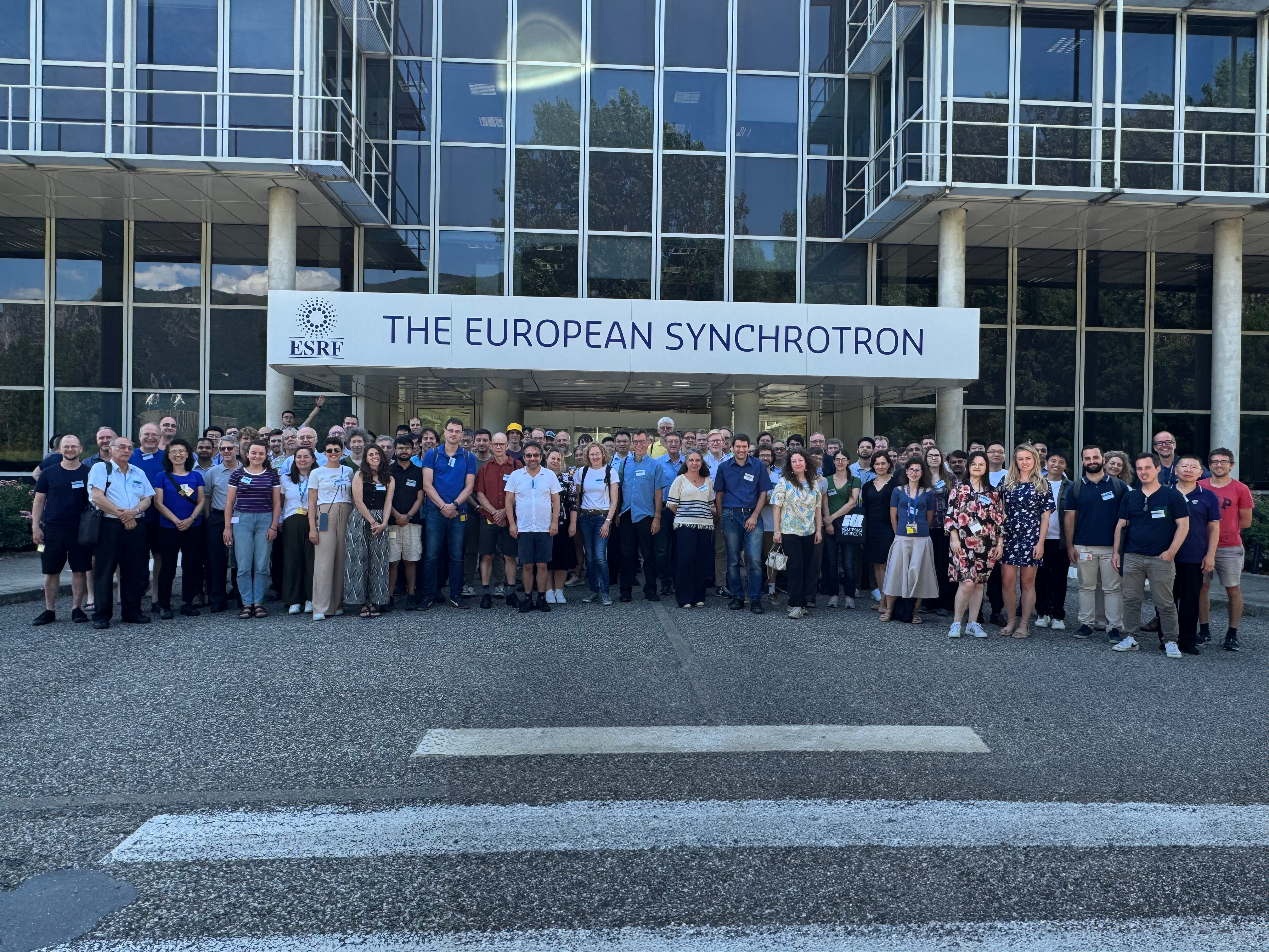
The 17th International Conference on Surface X-ray and Neutron Scattering (SXNS17) was held at the EPN campus in Grenoble, France, hosting the European Synchrotron Radiation Facility (ESRF) and the Institut Laue-Langevin (ILL) from July 15 -18, 2024.
The conference brings together researchers studying surfaces and interfaces in solid, liquid, biological, soft and hard condensed matter via neutron or X-ray (primarily using synchrotron sources) scattering techniques. Topics covered include instrumentation, studies of nanostructured surfaces and interfaces, surfaces and interfaces in soft matter, biological interfaces, magnetic thin films and interfaces, magnetic proximity effects, topological materials, emergent interfacial materials, and dynamics of surfaces, interfaces, and nanostructures.
The conference included the following topics:
Rare earth elements are utilized in a diverse range of modern and evolving technologies. Current methods for separating and purifying these elements involve their interactions at liquid interfaces. For example, the primary separations technique in current use, known as solvent extraction, involves molecular binding and assisted transport across liquid-liquid interfaces. Other techniques in development utilize rare earth element adsorption to liquid-vapor and liquid-solid interfaces. The development of these techniques has taken place largely in the absence of an understanding of interfacial distributions of rare earth elements, and of the binding, coordination, and ordering of rare-earth elements with molecular species at the interface. This provides an opportunity for the use of X-ray surface scattering and spectroscopy to investigate these issues. Hopefully, these investigations will inform the development of more efficient and cleaner separations processes of rare earth elements. In addition, the +3 charge on most rare earth ions and their complex coordination with many molecules provides an opportunity to understand new interfacial science. I will review X-ray studies in this area, mostly from my research group’s activities, with an emphasis on the information that can be learned from X-ray techniques that characterize rare earth elements at aqueous interfaces.
A precise understanding of the distribution of chemical species at interfaces, in particular ions, is crucial in many areas.
For example, the so-called Solid-Electrolyte Interface (SEI) plays a crucial role on battery performance.
In a different area, the interaction between ions, humics and minerals plays a key role in the dispersion of pollutants as the modulation of the effective surface charge by ion-surface interactions is affecting the structure of the electric double layer, hence the interaction between charged particles in aqueous solutions and floculation and precipitation.
Rare earth elements of high purity are critical for many industries and their recycling is essential to build a circular economy.
A widely used method for the separation of lanthanides is ion exchange - cation exchange with elution by complexing agents - In these processes, the order of elution of the different rare earth elements depends on the values of the stability constants of the complexes formed with a solid.
From a fundamental point of view, ions at the aqueous -solid interface also exhibit a range of fascinating phenomena, depending on the surface charge, ion valency, concentration, with sometimes extremely surprising and counter-intuitive behavior.
Looking at ionic distributions at the solid-aqueous interface offer a unique way to look at subtle effects related to polarization, hydration,... which would be very difficult to observe otherwise, with often very different behavior with ions with similarly charge and valency.
Based on previous work [1], we have developed a fully quantitative method that gives access to the detailed composition of the first Angstroms at the silica - electrolyte interface. Standing waves generated in a sub-micron electrolyte layer by Bragg reflection on W-Si multilayers with a 2nm period are used to excite fluorescence from the ions under consideration. Using a thin Cr reference layer in the multilayer, fully quantitative analysis of fluorescence intensity combined with reflectivity measurements allows us to precisely determine the electrolyte film thickness.
Several mixtures of rare earth chlorides and perchlorates were investigated, competitive adsorption being used to enhance differences between rare earth cations and increasing accuracy.
Adsorption is so strong that our results cannot be satisfactorily described by a classical Gouy-Chapman-Stern Model.
Instead, we observe a fairly good agreement with distribution profiles over the entire electrolyte layer based on original molecular dynamics simulations fully taking into account short and long range interactions [2].
Surprisingly, we observe up to 3 adsorption layers, explaining the strong adsorption and the failure of classical models.
[1] F. Malloggi et al., J. Phys. Chem. C 2019, 123, 30294−30304.
[2] B. Siboulet et al., J. Phys. Chem. C 2017, 121, 6756−6769.
Shear is observed in many natural and technological systems, affecting their structure, dynamics, function and performance. Entangled polymers exhibit unique flow behaviours, as relaxation processes occur on time scales relevant to our daily lives, from milliseconds to hours or even days. Investigating the relation between out-of-equilibrium microscopic structure and dynamics of fluids and their macroscopic rheological response can enhance our understanding of viscoelastic flow, leading to improved material properties and applications.
This study combines neutron reflectometry (NR), rheology, and computer simulations to characterize the behaviour of polystyrene (PS) brushes under shear by an entangled PS semi-dilute solution. Two brushes with different chain lengths and grafting densities were used. NR reveals similar shear effects on both brushes restricted to the overlap region, causing a decrease in brush thickness and a sharper brush-bulk interface. In addition, the brush thickness returns to equilibrium upon cessation of shear, and the effect can be cycled many times over. The collapse of the brush occurs regardless of the type of brush used, indicating that the dynamics governing the structural change are determined by the free chains in solution rather than the brush itself. Coarse-grained computer simulations of the interfaces were in agreement with the experimental data.
We have recently developed a novel setup that enables the characterisation of sheared interfaces by combined NR and polarised infrared spectroscopy. The main novelty of this setup lies in the use of polarised infrared spectroscopy, which allows following any anisotropy due to shear stress appearing at the interface and, thus, determines molecular orientation. Additionally, NR allows elucidating the structure of these brushes perpendicular to the interface. The first results obtained from this new setup will be presented.
This research shows the feasibility of engineering shear-responsive polymer brushes in entangled polymer solutions, with potential applications in nanosensors and dynamic surface friction and adhesion control.
References
[1] Wolf, M. et al. Combined neutron reflectometry and rheology. J. Appl. Crystallogr. 2013, 46, 1729-1733.
[2] Korolkovas, A. et al. Polymer Brush Collapse under Shear Flow. Macromolecules 2017, 50, 1215-1224.
Resonant x-ray scattering and diffraction is an emerging technique for the nanostructure characterization of soft matter. While these approaches are widely spread for highly ordered material, the amorphous or paracrystalline nature of most of the soft matter systems limits the use of currently developed method such as diffraction anomalous fine structure technique.
In this presentation, we will illustrate the use of resonant x-ray scattering on conjugated polymers used for optoelectronic devices. The performance of such devices are highly dependent upon the molecular packing however. The presence of sulfur atoms in common conjugated polymer such as P3HT or N2200 enable the use of tender x-rays (sulfur K-edge is around 2.47 keV) to probe and enhance signals from the sulfur atoms. Tracking the intensity variation across the sulfur K-edge and comparing with theoretical calculation enable to discriminate between different packing geometries and to extract important new microstructural information.
We will both present results in the transmission geometry, on thin films deposited on Si3N4 membranes, as well as in the grazing-incidence (GI) geometry on thin film on Si substrate. Especially for the GI geometry, we will discuss the importance of absorption and its impact/distortion on the collected signal.
[1] G. Freychet, et al., J. Am. Chem. Soc. 143, 1409 (2021).
[2] G. Freychet, et al., Mater. Horiz. 9, 1649 (2022).
Many innovative data analysis approaches have been developed since the inception of the reflectometry technique. Unfortunately many of them have fallen out of active use due to the lack of software tools implementing those approaches, that are available to the surface science community. In this talk we'll revisit several of those approaches and their application for studying polymer brush systems. The pivotal importance of tools in reproducible experimental workflows is explored, both from a toolmaker's point-of-view, and from that of a tool user.
Modern synchrotron beamlines and neutron instruments have undergone significant changes due to technological advances and newly deployed infrastructure. Thus, experiments are becoming more data-intensive and data-driven and increasingly relying on online data analysis for efficient use of experimental resources. In this regard machine-learning (ML) based approaches of specific importance for real-time decision-making based on online data analysis and connected closed loop feedback applications.
Following recent advances in ML-based analysis of x-ray reflectometry we present both the underlying ML models and concepts as well as the integration into closed loop operation in experiments.
Specifically, we present an approach that incorporates prior knowledge to regularize the training process across broader parameter spaces. This method proves effective in diverse scenarios relying on physics-inspired parameterization of the scattering length density profiles. By integrating prior knowledge, we enhance training dynamics and address the underdetermined (or "ill-posed") nature of the problem. We show that our approach scales well with increasing inverse problem complexity, performing efficiently for an N-layer periodic multilayer model with up to 17 open parameters.
Pithan et al., J. Synchrotron Rad. (2023). 30, 1064
Hinderhofer et al., J. Appl. Cryst. (2023). 56, 3
Munteanu et al., J. Appl. Cryst. (2024). 57, 456
Neutron reflectometry is a powerful tool for investigating thin films and interfaces, with a host of industrially relevant applications from model cell membranes to next generation hard drive materials and everything in between. The information which can be extracted has all the usual benefits of using neutrons: isotope and light element sensitivity, as well as absolute magnetometry, but is severely limited by neutron flux. After users have made unusually large samples, we still often have to cut the incident beam from cm down to 100um to keep the beam footprint solely on the sample.
This has been the standard modus operandi since the first reflectometry experiments approx. 40 years ago. However, since the realization of the Selene guide system (on AMOR at PSI), another option has existed – to focus the neutrons onto the sample, in our case directly from the moderator face. This approach gains orders of magnitude more flux than collimating but comes at a significant technical cost.
In this talk I give an overview of the ESTIA instrument at the ESS, highlight the challenges which come with trying to truly focus a neutron beam and go over some of the new experiments which will be possible.
Complementary to x-ray diffraction patterns that represent the crystal lattice in reciprocal space, the atomic pair distribution function (PDF) describes the structure of a material as a histogram of interatomic distances r in real space. The total scattering approach that enables PDF analysis requires that scattering data is collected over a wide Q range of the order of 20 Å-1 and subsequent Fourier transformation of the entire scattering pattern into real space. While total scattering at high-energy x-ray beamlines has become a standard technique for bulk-type samples, the unfavorable thickness ratio of a thin film (nanometer regime) to its substrate (micro- to millimeter regime) limits the detectability of the film signal in simple transmission geometry as described e.g. in Ref. [1]. For the hard x-ray range, grazing-incidence (GI) geometry is well established for surface scattering. GI diffraction and total scattering at high photon energies >50 keV are advantageous as they enable direct access to a wide Q range in a single exposure on a large area detector, but are more demanding in terms of experimental prerequisites. At beamline P07 at PETRA III (DESY, Hamburg), high-energy surface diffraction [2] and, more recently, total scattering in GI geometry [3,4] have been developed into routine capabilities that benefit various research fields from catalysis to electronics and strongly correlated materials. This presentation reports on the latest progress at PETRA III beamlines P07 and P21.1 in applying GI total x-ray scattering to study the short-range order in epitaxial layers at non-ambient conditions and in situ processes like phase formation during post-deposition crystallization and electrochemical thin film growth. Such novel insights into the evolution of atomic structure on short to long-range scale in thin film systems demonstrate the expected huge gain for communities that have so far lacked a tool like PDF analysis to determine the local structure and disorder in thin layers.
[1] K. M. Ø. Jensen et al., IUCrJ 2 (2015) 481-489.
[2] J. Gustafson et al., Science 343 (2014) 758-762.
[3] A.-C. Dippel, et al., IUCrJ 6 (2019) 290-298.
[4] A.-C. Dippel et al., Nanoscale 12 (2020) 13103-13112.
Neutron reflectometry (NR) is a powerful technique to explore the structure of the surface and interfaces of materials. In a typical NR experiment, the sample surface/interface must be uniform over an area of more than 10 cm2 due to the size of the neutron illumination. Therefore, conventional NR has been inapplicable for the structure analysis of the sample with in-plane inhomogeneity. We developed an NR imaging technique to examine the position-dependent NR profile. The spatially resolved NR profile perpendicular to the beam axis is directly measured with the sheet-like neutron illumination and a position-sensitive detector. An NR image is reconstructed by a computer tomography (CT) calculation for the reflectivity at a given momentum transfer, q, dependent on the in-plane rotation angle of the sample. The spatial resolution of the NR-CT method is determined by the collimation condition of the neutron beam and the resolution of the detector. With a neutron reflectometer SHARAKU in J-PARC, the achievable resolution is ca. 0.6 mm. The CT reconstruction images based on the reflectivity at various q provide the NR profile at each image pixel, and the depth structure of the neutron scattering length density can be evaluated at a local area as small as 0.1 mm2. In this presentation, the application of the NR-CT to the analysis of laterally inhomogeneous interfaces of a polymer system is also demonstrated.
This talk will present some comparisons of neutron and X-ray reflectivity of supported lipid members as platforms to investigate various membrane properties. I will emphasize some recent work on a robust polymer cushioned membrane system that recapitulates many salient features of the plasma membrane including control over the fluidity of the membrane and thickness/density of the underlying, pH responsive polymer network. The system is readily fabricated from commercially available materials with commonly available laboratory equipment. High quality, high coverage lipid membranes were constructed using Langmuir-Blodgett and vesicle fusion deposition methods. The underlying polymer network or membrane cushion is a covalently grafted PAA cushion with nominal thickness controlled by spin coating conditions. The pH-sensitive structure of the PAA network and coupling to the membrane can be used to control the hydrated thickness of the film and membrane diffusivity. At low pH, when the PAA is collapsed, diffusivity is strikingly lower than at high pH when the PAA is swollen. Under physiological conditions, the diffusion rates of lipid membranes on the PAA network were indistinguishable from those on bare glass supports. Importantly, the PAA cushioned membrane structure is stable during cycling through acidic, neutral, and alkaline conditions.
Polymer electrolyte fuel cells used in electric vehicles convert the chemical energy of hydrogen into electricity to power motors, in which the electricity is generated in three steps: hydrogen gas is dissolved into protons at the anode, the protons are transported through an electrolyte, and the protons are reacted with oxygen at the cathode. Perfluorosulfonic acids (PFSAs) are widely used as a proton conducting ionomers not only for the electrolyte but also for the binder of the carbon black with Pt catalyst in the cathode. Therefore, the proton conductivity and oxygen permeability of PFSA can be a bottleneck of the electrochemical reaction on the Pt surface at the cathode, which depend on the nanostructure as well as the chemical structure of PFSA. Industrially, the assembly of anode, electrolyte and cathode is made by the so-called decal process, in which the stack is hot-pressed at 120-140 °C for 10-20 minutes. The assembly is then combined into the fuel cells and annealed by the water produced by the reaction repeatedly over a run-in period. Although the processes are empirically tuned, the nanostructure of the PFSA is expected to be controlled to optimize the bottleneck.
In this study, the effect of the thermal annealing on a Nafion® thin film, typical PFSA, on a Pt coated Si substrate in the decal process is evaluated by grazing incident angle small angle x-ray scattering (GI-SAXS) mainly for in-plane nanostructure, neutron reflectometry (NR) for out-of-plane nanostructure, positron annihilation lifetime spectroscopy (PALS) for free volume, and x-ray absorption spectroscopy (XAS) and quartz crystal microbalance (QCM) for water content in a vacuum and in air, respectively, to understand the role of the decal process. Interestingly, the drastic change in the nanostructure was observed between 10 and 20 minutes, which is consistent with the annealing time of the decal process. Also, some of the experiments are performed with changing humidity with repetitions to evaluate the effect of solvent annealing on the run-in period.
We will show and discuss the results in detail in the presentation.
The use of an ionic liquid (IL) as electrolyte is promising for very high capacity energy storage units based on graphene electrode [1]. Moreover, introducing gold nanoparticles (NPs) at the interface between the IL and the electrode makes it possible to increase the capacitance [2]. We study the interface between these NPs and a layer of the [C20mim]+[NTf2]- IL. We use x-rays surface radiolysis [3] to produce gold NPs under a Langmuir film of [C20mim]+[NTf2]- deposited on an aqueous sub-phase containing gold ions [4]. The formation of gold NPs is obtained by irradiating the surface with the x-rays which simultaneously allows in following their growth and the structural transformations in the film. The films characterization was carried out on the liquid sub-phase by thermodynamic (surface pressure versus surface density isotherms), surface x-rays scattering measurements at the SOLEIL synchrotron and AFM measurements on films transferred on solid substrates before and after irradiation. We observe that both the film thickness and r, the gold ion/IL molecule ratio, are key factors for the formation of the gold NPs: at a low ratio (r = 30), gold NPs of about 15 nm in diameter are obtained. On the other hand, for a high ratio (r = 600), we observe the appearance of a superstructure in the monolayer but no formation of NPs. The exchange between AuCl4- (in the subphase) and +[NTf2]- appears to be the element preventing the possibility of forming gold NPs anchored under the layer [5].
References
[1] W. Raza et al , Nano Energy, 2018, 52, 441.
[2] M. Sarno et al, Journal of Physics and Chemistry of Solids, 2018, 120, 241.
[4] F. Muller et al, Langmuir, 2004, 20, 4791.
[5]G. Diot, PhD Thesis, Sorbonne Université 2023.
Polymer-surfactant (P-S) complexes have been a subject of extensive research efforts, due to their high industrial and scientific relevance both in solution [1] and on the interfaces [2]. A feature of polymer-surfactant complexes is that they are able to assemble into the supramolecular complexes above a certain critical association concentration (cac). However, our understanding of the intermolecular interactions underpinning the mechanism and pathway of the complex formation remains incomplete, especially in the case of structures formed in planar confinement.
In this work, in order to gain mechanistic insights, we have studied the interactions between a flexible polyelectrolyte and surfactants (amphoteric and anionic) in a weak interaction regime, where the driving force of the interaction is the van der Waals forces. Surface confined mesophase of these complexes were investigated using X-ray and neutron reflectivity, combined with grazing incidence diffraction. We have found spontaneously formed highly porous multilayer structures (Figure 1, A), facilitated by the presence of the polymer, contrary to the rupture of multilayer vesicles, observed earlier [3]. Formed films are responsive to humidity, which promoted reorganization of the multilayer and melting of the surfactant crystal, formed in the spin-coating process, in favour of formation of the liquid-crystal-like structure with correlated roughness, as indicated by the diffuse scattering (Figure 1, B)
Our results show spontaneous formation of structured film during both spin- and dip-coating of the complexes. We have established that surfactants are uniformly distributed in the polymer film and reorganize upon exposure to humidity and investigated the influence of complex formation in solution. Finally, we have compared the structure of surface confined mesophase with the precipitates formed when the electrostatic attraction dominates.
Figure 1. A. Neutron reflectivity profiles for hydrogenated and deuterated surfactant contrast, measured at 0% humidity (dry) and 80% humidity (wet). B. Reciprocal space map obtained during X-ray reflectivity measurement of the same composition sample at 60% humidity. Note the presence of periodic diffuse scattering stretched along qx direction, indicating roughness correlations in the multilayer.
[1] C. D. Bain, et al., Complexes of surfactants with oppositely charged polymers at surfaces and in bulk. Adv.Col.Int.Sci, 155(1):32–49, 2010.
[2] L. Fernandez-Pena, et al., Impact of the bulk aggregation on the adsorption of oppositely charged polyelectrolyte-surfactant mixtures onto solid surfaces. Adv.Col.Int.Sci, 282:102203, 2020
[3] Micciulla, S. et al. One-step procedure for the preparation of functional polysaccharide/fatty acid multilayered coatings. Commun Chem 2, 61 (2019).
Understanding and controlling structure and function of liquid interfaces is a constant challenge in biology, nanoscience and nanotechnology, with applications ranging from molecular electronics to controlled drug release. X-ray reflectivity and grazing incidence diffraction provide invaluable probes for studying the atomic scale structure at liquid–vapour interfaces. The new time resolved laser system at the LISA liquid diffractometer [1] situated at beamline P08 [2] at the PETRA III synchrotron radiation source in Hamburg, Germany provides a laser pump - X-ray probe setup to study dynamics at the liquid interface. The femtosecond laser combined with the LISA diffractometer allows unique opportunities to investigate photo-induced structural changes at liquid interfaces on the pico- and nanosecond time scales with pump-probe techniques. The synchronisation of X-ray and laser pulses (see Figure 1) enables to achieve a time resolution of 80 ps, which was verified with Bismuth. First experiments include laser induced effects at salt solution and liquid mercury surfaces with static and measurements on varied timescales showing the proof of concept for investigations at liquid surfaces. [3]
References
[1] B. M. Murphy et al., Journal of synchrotron radiation 21, 45-56 (2024).
[2] O. Seeck et al., Journal of synchrotron radiation 19, 30-38 (2012).
[3] J. E. Warias et al., Journal of synchrotron radiation 31, 779-790 (2024).
[4] F. Reise et al., Chemistry – A European Journal 24, 17497-17505 (2018).
The horizontal time-of-flight neutron reflectometer at the reactor of the Delft University of Technology, The Netherlands, has been completely renewed, relocated, and upgraded and allows for the study of air/liquid, solid/liquid, and solid/air interfaces and is now accesible for researchers from around the globe. Innovations in the redesign include (i) a completely flexible double disk chopper system allowing to choose the optimal wavelength resolution with exchangeable neutron guide sections between the chopper disks to increase intensity, (ii) a movable second diaphragm just before the sample position to better control the beam footprint on the sample and effectively decrease counting times, and (iii) guides along the entire flight path of the neutron reflectometer. The performance of the renewed reflectometer is illustrated with measurements of hydrogen sensing materials, and a comparison will be made before and after the installation of the cold neutron source.
In this study, we introduce cutting-edge neural network algorithms that precisely predict crystal unit cell parameters and contact planes from Grazing Incidence X-Ray Diffraction (GIXD) data. Our method processes a list of q-positions and delivers predictions of the unit cell with exceptional accuracy—better than 0.1 Angstrom in dimensional precision and sub-degree in angular measurements. It eliminates the necessity to measure the 'missing wedge' in a specular scan, simplifying the experimental setup. Our AI method facilitates the rapid, autonomous processing of complex GIXD patterns without user intervention. It enables a detailed evaluation of the analysis's sensitivity to missing or spurious peaks due to its ability to predict structures for a large number of GIXD patterns quickly. Indeed the analysis copes well with patterns that miss peaks or contain only few peaks in total. These advancements present a substantial improvement in efficiency and reliability for researchers utilizing intricate GIXD patterns in crystallographic surface science studies.
With the advancing of large-scale integrated circuits, the sizes of element devices have shrunk to nanoscale. At this size scale, quantum effects emerge and dominate the performance of the low-dimension materials and devices. To study the low-dimension structures and their dynamics based on coherent surface x-ray scattering (SXRS), we are constructing the Low-Dimension Structure Probe (LoDiSP) beamline at High Energy Photon Source (HEPS). It uses an in-vacuum undulator as the photon source, and connect the Multi-Environment X-ray Scattering station (EH1) and the In-situ Coherent Surface X-ray Scattering station (EH2) in tandem. At EH1, a large-load 6+3-circle Huber diffractometer equipped with various sample environment setups provides various x-ray scattering/diffraction methods. At EH2, a “hybrid MBE+SXRS+RHEED+XPCS” integrated facility enables in-situ growth of samples in ultra-high vacuum chamber and coherent SXRS characterization. An evanescent waves modulator will be developed to artificially transform the growth mode of multi-element compound thin films and be combined with grazing incident x-ray photon correlation spectroscopy (GI-XPCS) to investigate the surface dynamics of thin films.
Neutron reflectometry (NR) is intrinsically a low-resolution structural technique, yielding one-dimensional scattering length density profiles that are often inadequate for detailed structural biology applications. However, NR stands out as one of the few methods capable of characterizing membrane-bound proteins and protein assemblies in a biomimetic, solvent-immersed, and thermodynamically relevant state, making it invaluable for biophysical studies. Integrating NR with complementary experimental data and molecular simulations can overcome its resolution limitations and obtain detailed three-dimensional structural information.
In this presentation, I will discuss our recent advancements in applying this integrative approach to study the homodimer formation of HIV-1 Nef at the lipid membrane. HIV-1 Nef is crucial for viral infectivity, high-titer replication in vivo, and immune evasion in HIV-infected cells. Nef functions not through intrinsic biochemical activity but through interactions with various host cell signaling proteins and intracellular trafficking pathways. Previous studies have underscored the importance of Nef homodimer formation at the plasma membrane for its functions.
In collaboration with the Smithgall group at the University of Pennsylvania School of Medicine, we combine NR of full-length myristoylated Nef bound to model lipid membranes with molecular simulations of high-resolution Nef monomer and homodimer structures [1]. This integrated approach provides direct evidence that Nef associates with the membrane as a homodimer, with its structured core region positioned away from the membrane to engage partner proteins. Additionally, studies on a dimerization-defective mutant, Nef-L112D, reveal that the helical dimerization interface seen in previous crystal structures is crucial for stabilizing the membrane-bound dimer.
[1] Heinrich F., Thomas E. C., Alvarado J. J., Eells R., Thomas A., Doucet M., Whitlach K. N., Aryal M., Lösche M., Smithgall T. E., Neutron Reflectometry and Molecular Simulations Demonstrate HIV-1 Nef Homodimer Formation on Model Lipid Bilayers. Journal of Molecular Biology 435(8):168009 (2023), DOI: 10.1016/j.jmb.2023.168009
Lipid membranes are the backbone for biologicals cells and to understand intercellular, and cell-cell interaction is of fundamental interest. The lipid molecules within the membrane define the cell properties and mesophase, and influence the functionality and efficiency of membrane proteins. [1,2,3] Therefore, controlled interaction between proteins and lipids in the biological membrane yields high potential for efficient drug transportation and release of pharmaceuticals. We want to present a model system consisting of DPPC and photoswitchable azobenzene amphiphiles [4] that induced structural changes of the membrane morphology upon isomerisation of the azobenzene mimetics by illumination with 365 nm and 455 nm to switch between the cis and trans isomer. In-situ X-ray and neutron reflectivity studies on Langmuir monolayers containing 5% [5] and 10% azobenzene amphiphiles at the water-air interface revealed an additional phase transition and light induced bidirectional, repeatable and fully reversible structural changes of the layer thickness and surface pressure. Further studies on liquid crystals containing DPPC and different percentages of azobenzene amphiphiles showed light-induced mesophase transitions between multilamellar (MLV) and bicontinuous cubic Pn3m structure at room temperature as visualised exemplarily in Figure 1 [6]. Recent times resolved SAXS measurements allowed to investigate the kinetics and dynamics of the mesophase transition for liquid crystals in dependence of the chemical composition of different azobenzene amphiphiles and identify transition times of up to several minutes. In addition to the scientific results we will also present the FAIR data handling and usage of data repositories and sample identifiers following the FAIR standards proposed by DAPHNE4NFDI [7] for an example SAXS data set.
Figure 1. Small angle X-ray scattering data of mixed DPPC and azobenzene mimetics for the cis (upper curve) and trans (lower curve) state and the fit (in grey) for the mesophases shown schematically on the right. [6]
[1] Cournia, Z., Allen, T. W., Andricioaei, I., Antonny, B., Baum, D., Brannigan, G., Buchete, N.-V., Deckman, J. T., Delemotte, L., Del Val, C., Friedman, R., Gkeka, P., Hege, H.-C., Hénin, J., Kasimova, M. A., Kolocouris, A., Klein, M. L., Khalid, S., Lemieux, M. J., Lindow, N., Roy, M., Selent, J., Tarek, M., Tofoleanu, F., Vanni, S., Urban, S., Wales, D. J., Smith, J. C. & Bondar, A.-N. (2015). The Journal of membrane biology, 248, 611.
[2] Shah, J. C., Sadhale, Y. & Chilukuri, D. M. (2001). Advanced Drug Delivery Reviews. 47, 229.
[3]. Phillips, R., Ursell, T., Wiggins, P., Sens, P. (2009). Nature 459, 379.
[4] Reise, F., Warias, J. E., Chatterjee, K., Krekiehn, N. R., Magnussen, O., Murphy, B. M., Lindhorst, T. K. (2018). Chemistry – A European Journal 24, 17497-17505.
[5] Warias, J. E., Reise, F., Hövelmann, S. C., Giri, R. P., Röhrl, M., Kuhn, J., Jacobsen, M., Chatterjee, Arnold, T., Shen, C., Festersen, S., Sartori, A., Jordt, P., Magnussen, O., Lindhorst, T. K., Murphy, B. M. (2023). Scientific reports, 13, 11480.
[6] Hövelmann, S. C., Dieball, E., Kuhn, J., Dargasz, M., Giri, R. P., Reise, F., Paulus, M., O., Lindhorst, T. K., Murphy, B. M. submitted.
[7] DAPHNE4NFDI is a National Research Data Infrastructure consortium funded by the German German Research Foundation (DFG). https://www.daphne4nfdi.de/
Mitochondria are well known as the ‘powerhouse of the cell’. Mitochondria also play a crucial role in apoptosis, a programmed mechanism of cell death. This process is tightly regulated by proteins at the mitochondrial outer membrane (MOM).
In this mechanism of cell death, the pro-apoptotic Bax protein is attracted to the outer mitichondrial membrane (MOM), where it induces membrane leakage. [1] In healthy cells, Bax is neutralized by the anti-apoptotic Bcl-2 residing in the MOM.[2]
The complexity of the MOM system makes it difficult to obtain an overall picture of its organisation. However, preparation of planar lipid bilayers and analysis with neutron reflectivity (NR) and attenuated total reflection- fourier transform infrared spectroscopy (ATR-FTIR) can be used to identify the position of the two crucial proteins in respect to the membrane and to identify the kinetics of the interaction. We can characterise this biological effect by employing hydrogen/deuterium labeling to create a stong contrast between the lipid and protein components.
Our results provide the first structural evidence of Bcl-2 preventing membrane perforation by Bax as part of its anti-apoptotic mechanism. In the absence of Bcl-2, the Bax protein both inserts into the lipid bilayer and removes lipids, forming a lipid-protein complex on top of the original bilayer structure. However, when Bcl-2 is reconstituted into POPC bilayers, Bax associates to the bilayer but does not insert into it or remove lipids. Separately when Bcl-2 is reconstituted into 9:1 POPC: cardiolipin bilayers (a higher cardiolipin content than the average MOM), Bax still perforates the bilayer with slower kinetics than in the absence of Bcl-2.[3] These results indicate that membrane lipids play an important role in apoptotic pore formation, and life hangs in the balance of membrane protein content and lipid composition.
[1] M. Lidman, BBA-Biomembranes 1858, 1288-1297 (2016).
[2] A. Mushtaq, Commun Biol 4, 507 (2021).
[3] L. A. Clifton, L.A., Science Advances, 9,22, (2023)
Deoxyribonucleic acid (DNA) is a negatively charged bio-macromolecule that helps in the transmission of genetic information for the growth and functioning of a living organism. Therefore, it is considered as a potential tool in gene therapeutics. Packing or condensing of this macromolecule is difficult because of the intra and intermolecular repulsive electrostatic and entropic interactions. Even though there are reports of condensing the molecule using inorganic salts in bulk aqueous medium, the assembly at the air-water interface is rarely reported. Here, we report the assembly of the DNA molecule at the interface induced by an imidazolium based ionic liquid (IL) 1,3 didecyl-2-methylimidazolium chloride. The surface pressure-area isotherm ensures the presence of the molecule at the interface with a high mean molecular area. Interfacial rheology measurements quantify the elastic nature of the molecular film. The storage and loss modulus of the film is found to strongly depend on the in-plane pressure. Advanced in-situ synchrotron X-ray reflectivity (XRR) study relates these physical properties of the film with its structure. The electron density profile of the film across the interface manifests the compact nature of the film in presence of the IL. This work suggests an easy way of immobilizing the DNA macromolecule at the air-water interface. The work has been extended to bulk aqueous solution of DNA in presence of the organic salt. Small angle X-ray scattering (SAXS) results indicate rigidification of the DNA strands in presence of the IL which is opposite of what is reported for an inorganic salt. Computational analysis of the work has provided the molecular details of this exceptional behavior in presence of the organic salt.
Plasma proteins are often used as biomarkers for severe pathologies including cancers and autoimmune diseases since they provide a circulating representation of the body conditions. Nevertheless, their detection in blood samples is still challenging due to their low abundance (pico- and nano-grams level), but mostly due to their glycosylated state which is believed to favor protein-lipid complexes. In this context, the study and understanding of protein-lipid interactions is of great importance, especially to optimise the sensitivity and reliability of biosensing techniques to identify protein biomarkers, in particular when present in their glycosylated forms [1].
In the project we investigated at nanostructural level the interaction of glycosylated protein biomarkers with biologically relevant lipid bilayer membranes using neutron and x-ray reflectometry, to identify how different lipids and glycosylation can affect this interaction to build a potential biosensor that is aimed for early detection of scarce biomarkers in blood samples.
The glycosylated proteins of interest for the study are soluble vascular-endothelial cadherin (sVE) and alpha-fetoprotein, two clinical biomarkers found in the blood for the detection of vascular abnormalities and liver cancer respectively [2,3,4].
Neutron and x-ray reflectivity results, together with QCM-D complementary data, showed significant changes in the lipid bilayer after the injection of glycosylated proteins, while smaller changes were reported in presence of non-glycosylated protein. We highlighted that the kinetic has an important role for the interaction and we proved that the interaction behavior is influenced by the lipid composition of the system as well as the degree of glycosylation on the protein.
Thanks to the study and glycosylation chemistry, it was possible to set the basis for the development of a lipid-based protein-biosensor in collaboration with the industrial partner of the project.
References
[1] A. Khalil-Mgharbel, et al. Biotechnology Journal (2018), 13:1800463.
[2] Blaise, S., et al. Vasc. Med. 20, 557–65 (2015)
[3] T. Kondo, Expert Rev. Proteom. (2014), 11, 9
[4] A. Alyass et al., BMC Med. Genom. (2015), 8, 33
Invasive fungal infections (IFIs) are an underappreciated public health threat that cause > 1.6 million deaths p/a with mortality rates between 20-80% [1,2]. Amphotericin B (AmB) is one of the most effective anti-fungal drugs. It kills fungal cells by binding to ergosterol in fungal cell membranes, causing membrane permeabilisation and changes in ergosterol-dependent signalling pathways [3 4]. Unfortunately, the structural similarity between ergosterol and cholesterol (mammalian cells) means AmB shows poor selectivity, resulting in dose-limiting toxicity causing severe side effects. Despite this, AmB remains a life-saving, last-resort drug because of its potent, broad-spectrum activity and low resistance rates [3]. There is a need to develop AmB-based treatments with reduced toxicity.
Recent work has revealed that the peptide Lactofungin (LFG) is synergistic with AmB. [5,6] LFG reduces the AmB dose 4 to 16-fold for many clinically relevant fungal pathogens. Recent work (Deplazes, unpublished) shows that LFG is non-toxic to human liver and kidney cells and non-haemolytic. This makes LFG a promising lead compound for developing an adjuvant drug that reduces the toxicity of AmB. We have shown that the synergy observed in cells can be reproduced in sterol-containing phospholipid bilayers, indicating the synergy is likely to be lipid dependent. Therefore model membrane systems can be used to elucidate the yet unknown mechanism of synergy.
Previous neutron reflectomtery experiments [7] have shown that at high concentrations AmB inserts into the lipid chain region of both pure POPC and sterol-containing membranes and does not significantly perturb the structure of pure POPC membranes. Furthermore AmB extracted ergosterol but not cholesterol, inserting more so into cholesterol-containing membranes. Our approach has been, initially, to reduce the AmB concentration 40 fold, closer to the clinically relevant dose and study the interactions with sterol containing membranes via a number of techniques including neutron reflectometry. Electrical impedance spectroscopy showed that LFG increases the AmB-induced disruption in an ergosterol-selective manner. Ultraviolet-visible spectroscopy and isothermal calorimetry show that, in contrast to Bovine Serum Albumin, the peptide LFG has negligible membrane binding and does not alter the solubility of AmB. This presentation will focus on our reflectivity work and outline future prospects.
References:
1. Fisher, M. C. & Denning, D. W. Nature Reviews Microbiology, 1-2 (2023).
2. Boroujeni, Z. B. et al. Microbial pathogenesis 152, 104616 (2021).
3. Cavassin, F. B. et al. Infectious Diseases and Therapy 10, 115-147 (2021)
4. Kamiński, D. M. European biophysics journal 43, 453-467 (2014).
5. Fernandes, K. E. & Carter, D. A. Frontiers in microbiology 8, 2 (2017).
6. Fernandes, K. E., Payne, R. J. & Carter, D. A. Antimicrobial Agents and Chemotherapy 64, e00842-00820 (2020).
7. Delhom R., Nelson A., Laux L., Haertlein M., Knecht W., Fragneto G. & Wacklin-Knecht H.P., nanomaterials, 2020, 10, 2439.
Dynamic surfaces and interfaces in batteries
Understanding the dynamic properties of surfaces and interfaces is foundational to many scientific disciplines. In electrochemistry, charge transfer occurs at the solid–liquid interface between electrode and electrolyte. For almost all systems and under most conditions, these interfaces are dynamic and evolve with time. This is particularly prominent for the surface electrochemistry in ion batteries because these are operated outside the electrochemical stability window of typical aprotic electrolytes leading to the formation and evolution of the so-called solid electrolyte interphase (SEI) [1,2]. Via its electron-blocking and ion-conduction properties, the SEI governs battery cell lifetime and kinetics [1].
In the first part of the talk, the particular usefulness of X-ray methods for studying electrochemical systems in general and the surface electrochemistry of ion batteries in particular will be introduced. I.e., X-ray methods can be performed in operando modality, and they provide quantitative information on an absolute scale (“X-ray Coulomb counting”).
The second part of the talk will focus on several examples in which we utilized surface X-ray scattering to understand the surface electrochemistry of ion batteries using model electrodes. The examples include the growth and evolution of the SEI on Si anodes and the respective correlation between SEI structure and electrochemical performance [3]. Moreover, the origin of LiF in the SEI will be addressed. Towards this end, we sought out a multimodal operando (synchrotron X-ray-based) experimental and theoretical approach [4]. Our results reveal that LiF nucleates via the electrocatalytic reduction of HF followed by significant PF6- anion reduction. Furthermore, X-ray chemistry-X-ray probe experiments of super-concentrated “water-in-salt” electrolytes will be discussed, in which we observed interfacial speciation-dependent surface-reduction of anions [5].
The final part of the talk will be devoted to future science-driven opportunities for surface X-ray scattering at DLRs and XFELs under the mottos “Surface X-ray scattering goes fast”, “Surface X-ray scattering goes ultrafast”, and “Surface X-ray scattering goes small”.
[1] Xu, J. Power Sources 559, 232652 (2023)
[2] Cao, Steinrück, Molecular-scale synchrotron X-ray investigations of solid-liquid interfaces in lithium-ion batteries, Encyclopedia of Solid-Liquid Interfaces (2024)
[3] Cao et al., Acc. Chem. Res. 52, 2673-2683 (2019)
[4] Cao et al., Chem. Mater. 33, 7315-7336 (2021
[5] Steinrück et al., Angew. Chem. Int. Ed. 59, 23180-23187 (2020)
Fluid interfaces with adsorbed substances are present in many systems in nature and industrial processes. Such interfaces often have a complex structural configuration which confers them the capability to withstand deformations. Consequently, they have been the object of study in recent decades both from a structural and rheological point of view. To date, there have been limited examples of simultaneous measurements of the interfacial rheology and the structure of complex fluid interfaces. This aspect holds particular significance, considering the challenge of comparing independently conducted structural and rheological experiments, where reproducing identical experimental conditions, such as temperature and/or concentration and/or compression history for monolayer at the air-water interface, is difficult. Specifically, there is great interest in the study of Langmuir monolayers of fatty acids and phospholipids which appear in many biophysical processes. This work focuses on the development, building, and exploitation of an interfacial shear rheometer, with DWR geometry, to be used on the neutron horizontal reflectometer FIGARO at Institut Laue-Langevin (ILL). Consequently, the instrument allows for simultaneous measurements of neutron reflectometry and interfacial rheology. In particular, a DWR probe 3D printed in titanium has been commissioned for the Anton Paar MCR702e Space rheometer available at the PSCM, and a suitable shear channel with annular geometry (machined in PTFE) has been designed and built to be used in the Langmuir trough. Notably, an improved Flow Field-Based data analysis software package has been developed to properly subtract bulk phases contribution, taking into account non-linear velocity profiles. The performance of the new instrument is illustrated with a study of the isothermal compression of C19 fatty acid Langmuir monolayers (FALMs). Additionally, Brewster Angle Microscopy (BAM) has been used to observe the formation of structures above the micron scale at the interface. The studies carried out attempt to shed light on the mechanism of loss of molecules observed in condensed phases at high interfacial pressures in monolayers of fatty acids from a dynamic and structural point of view.
Transport phenomena at interfaces include amongst others the transport of heat or charges, which are of eminent importance in electronic or energy harvesting devices [1]. By using pulsed x-ray scattering with 80 ps time resolution following laser excitation of thin films [2] we were able to resolve the caloric balance in layered structures of gold and a skutterudite, CoSb$_3$. From the dynamics of heat distribution we conclude on the importance of energy transport across the gold-CoSb$_3$ interface, which is to a large part carried not by phonons, but by the heated electron gas. Gold is known as a good source for injecting electrons in coupled subsystems due to its weak electron-phonon coupling. The amount of heat transfer across the interface suggests additional transport by ballistic electrons [3].
[1] H. Bracht, S. Eon, R. Frieling, A. Plech, D. Issenmann, D. Wolf, J. Lundsgaard Hansen, A. Nylandsted Larsen, J. W. Ager III, E. E. Haller, New J. Phys. 16, 015021 (2014)
[2] A. Plech, B. Krause, T. Baumbach, M. Zakharova, S. Eon, C. Girmen, G. Buth and H. Bracht, nanomaterials 9, 501 (2019).
[3] A. Plech, P. Gaal, D. Schmidt, M. Levantino, M. Daniel, S. Stankov, G. Buth and M. Albrecht, New J. Phys. (2024) submitted.
The modification of epitaxial layers through hydrogenation is a thriving field of research that offers diverse opportunities to tune the physical properties of different systems. Recent research has been extended to correlated oxide interfaces, where hydrogen induced, reversible metal-to-insulator transitions have been uncovered in material systems such as rare-earth nickelates [1-2]. The combination of neutron and x-ray scattering methods are without doubt one of the best way to study these systems. In this talk we will show the reentrant phase transformation observed at room temperature for LaNiO$_{3}$ upon exposure to hydrogen gas. Electrical transport shows a metal-insulator-metal transition which was never observed before. We will present the results of in situ neutron reflectometry (NR), which allowed us to distinguish and quantify oxygen depletion and hydrogen incorporation, which are two possible mechanisms to explain the electronic modification of the host layer. In addition to the neutron measurements, we will present complementary results from in situ synchrotron x-ray diffraction and x-ray absorption, which helped to uncover the hydrogenation mechanism and electronic modification of the Ni valence. This work clearly shows the need of combining both neutron and x-ray scattering techniques to correctly identify the source of modifications induced by gas exposure in complex systems such as transition metal oxides.
References
[1] J. Shi, Y. Zhou, S. Ramanathan, Nat. Commun. 2014, 5, 4860.
[2] Haowen Chen, et al. Nano Letters 2022 22 (22), 8983-8990
Surface acoustic wave (SAW) devices are key components for processing radio frequency signals in wireless communication, known for their high performance, compact size and low cost. A quantitative understanding of energy conversion and loss mechanisms is essential to improving device structures and materials. Using the 100 ps bunch length of The European Synchrotron ESRF, our stroboscopic full-field diffraction x-ray microscopy (s-FFDXM) studies of a prototypical one-port resonator device reveals multiple sources of acoustic loss. The exceptional strain sensitivity allows measurement of acoustic waves with picometer-scale amplitude. A difference in the apparent resonant frequencies observed in the x-ray and electrical measurements results from the spatial non-uniformity of the acoustic excitation and from substantial leakage out of the active area. The high-resolution spatiotemporal imaging demonstrated in this work is generally suited for studying nano-phononics, specifically when the feature size is smaller than the optical wavelength. Applying s-FFDXM and an associated wave decomposition analysis method in combination, we achieve high-resolution spatio-temporal imaging of the acoustically induced strain waves. The method has a strain sensitivity of ∼ 10−7, corresponding to a surface displacement on the order of 1 pm. Measurements on a prototypical one-port SAW resonator reveal multiple mechanisms of acoustic loss with distinct spatial and time dependencies. Specifically, the maximum acoustic energy in the cavity area was not observed at the intended resonance frequency despite a well-matched IDT period and a higher reflector efficiency. A substantial amount of acoustic energy was leaked to the side (22%) and through the reflector (13%), leading to a shift in the resonance frequency of –9 MHz.
Liquid surfaces and interface abound in living matter, industrial processes, and everyday life and activities. Elucidating their structure is prerequisite to the understanding of the functionalities involved and thus highly important. This talk will address briefly some of the liquid surfaces and interfaces studied to date by x-ray methods, e.g. “simple” liquids like water [1] and liquid metals [2], and the more intricate chain molecules [3]. The main focus will be, however, on Room Temperature Ionic Liquids (RTILs) [4,5], a novel class of Complex Liquids, the study of which has grown explosively since the mid-1990’s, due to their many applications, ranging from electrochemical devices, through lubrication and pharmaceutical synthesis, to environmental application like CO2 capture and industrial sludge treatment, and even to targeted drug delivery within the human body. Their tunable balance of competing dominant Coulomb and Van der Waals interactions, along with a number of more-minor other interactions, renders RTILS most interesting scientifically as well. We will discuss our recent x-ray studies of the surface and bulk structure of a model homologous series of RTILs, [CnMIM][NTf2], (n denoting the number of carbons in the cationic alkyl chain), which reveal the surface-bulk structural relations, and demonstrate intriguing structural variations with n, temperature, and the nature of the interface.
[1] A. Braslau et al. PRL 54, 114(1985); M. Fukuto et al. PRL 117, 256102(2016).
[2] O.M. Magnussen et al. PRL 74, 4444(1995); O. Shpyrko et al. PRB 69, 245423(2004); S. Mechler et al. PRL 105,186101(2010).
[3] X.Z. Wu et al. Science 261, 1018 (1993); B.M. Ocko et al. PRE 55,3164 (1997); S. Guttman et al. PNAS 113, 493(2016); S. Das et al. Nat. Phys. 18, 1177(2022)
[4] E. Sloutskin et al., JACS 127, 7796(2005); M. Mezger et al. Science 322, 428(2008); PNAS 110, 3733(2013).
[5] J. Haddad et al. PNAS 115, E1100(2018): D. Pontoni et al. SM 13, 6947(2017); JPCC 123, 3058(2019); JML 355, 118874 (2022); JCIS 661, 3(2024).
The need for sustainable energy, reduction of pollutants, and the environmental benign processing of chemicals has spurred worldwide scientific activities in electrochemical energy science and electrocatalysis. These processes occur at the interfaces of solid catalyst materials in contact with complex liquid environments and under conditions involving high reaction rates, pronounced mass transport, and vigorous gas evolution. Because conventional surface analytic techniques cannot directly access these interfaces, the atomic scale surface structure and composition of the catalysts controlling the reactions are notoriously difficult to determine. For a better understanding of structure-property relationships and catalyst degradation mechanisms, experimental approaches are required that provide insight into the atomic and nanoscale interface structure in operando under reaction conditions. We have developed high-energy X-ray scattering methods for in situ and in operando studies of the complex interfaces between electrocatalysts and liquid electrolytes under reaction conditions. These methods allows to obtain very large datasets in short time, from which the interface structure can be determined with unprecedented detail, as demonstrated here by studies of the atomic-scale surface structure of Pt(111), Pt(110), and Pt(100) during oxide formation and reduction in perchloric and sulfuric acid solution. The obtained results on the potential-dependent oxide structure provide insights into the oxidation-induced Pt degradation by Pt dissolution and surface restructuring.
The presence of specifically adsorbed anions can significantly affect the electrochemical reactivity of a metal electrode which is of major interest for galvanic deposition, etching, corrosion and electrocatalysis. In-situ surface x-ray diffraction has enabled an atomic/molecular-level understanding of the interface under reactive conditions, including its potential and time dependence, to be developed. While information about the atomic structure of the electrode surface in electrochemical in-situ cells has been widely investigated, insight into the charge distribution and the structure of the electrolyte at the interface is still lacking. Advances in these directions offer possibilities in elucidating atomic scale models of the electrochemical interface and thus will help to establish structure-stability-reactivity relationships.
A fundamental understanding of the nature of the charge transfer, especially the influence of the applied potential and the screening by the electrolyte, is a major goal in electrochemistry to better understand electrochemical processes and charge transfer during adsorption and deposition. [1]
Thus combining x-ray spectroscopy and x-ray diffraction to gain site specific information about the charge distribution at buried interfaces is a promising tool. [2,3]
Examples of how the use of surface x-ray scattering techniques can help to characterise electrochemical interfaces in-situ in order to link, structure, reactivity and stability will be presented. [4-5] Advances in these directions offer possibilities in elucidating atomic scale models of the electrochemical interface and thus will help to establish structure-stability-reactivity relationships and to understand growth kinetics and electrochemical phase formation.
References:
[1] Y. Gründer and C. A. Lucas, Nano Energy 29, 378 (2016).
[2] Y. Gründer, P. Thompson, A. Brownrigg, M. Darlington, and C. A. Lucas, Journal of Physical Chemistry C 116, 6283 (2012).
[3] Y. Joly et al., Journal of Chemical Theory and Computation 14, 973 (2018).
[4] Y. Grunder et al., Charge Reorganization at the Adsorbate Covered Electrode Surface Probed through in Situ Resonant X-ray Diffraction Combined with ab Initio Modeling; Phys. Chem. C 2022, 126, 9, 4612–4619
[5] Yvonne Soldo-Olivier et al., Unraveling the Charge Distribution at the Metal-Electrolyte Interface Coupling in Situ Surface Resonant X-Ray Diffraction with Ab Initio Calculations, ACS Catal. 2022, 12, 4, 2375–2380
Responsive nanostructured films from amphiphilic lipids hold significant potential for applications in food, pharmaceutical, and biotechnological sciences. For instance, the amphiphilic lipid glycerol monooleate can be used to form antimicrobial coatings [1,2]. Lyotropic liquid crystalline films based on glycerol monooleate are also interesting as nanoreaction environments, drug delivery platforms, and biosensors. However, the nanoscale structural changes and orientations that such films undergo on surfaces during changes in composition or chemical reactions are not yet fully understood.
This presentation discusses the creation and analysis of mesostructured lipid films. We utilize in situ time-resolved grazing incidence small-angle X-ray scattering (GISAXS) to investigate the formation and transformation of film structure, orientation on the surface, and dynamics upon variation in composition and during chemical reactions. We demonstrate the formation of food-grade glycerol monooleate/triolein coatings on glass and silicon wafers without prior surface modification, employing ethanol as a coating solvent to achieve ultra-low contact angles [3]. Using a GISAXS sample platform, we examine the effects of composition, humidity, drug loading, and chemical reactions on the coating nanostructure. Our latest results, combining in-situ GISAXS with ellipsometry and confocal Raman microscopy studies, show the enzymatic hydrolysis of glycerol-lipids in aqueous enzymatic solutions, leading to swelling of the internal film nanostructure and eventual structural transformations [4]. Additionally, we analyze novel enzyme-responsive coatings and nano-reaction platforms to study polymerization reactions [5]. These findings highlight the role and application of lipid self-assembly in thin films for the functional modification of various substrates.
References:
[1] Stefan Salentinig, Mahsa Zabara, Pietro Parisse, and Heinz Amenitsch, Phys. Chem. Chem. Phys., 2018, 20, 21903-21909
[2] Mahsa Zabara, Qun Ren, Heinz Amenitsch, and Stefan Salentinig, ACS Applied Bio Materials, 2021, 4, 5295-5303
[3] Stefan Salentinig, Mahsa Zabara, WO/2020/035483 - ANTIMICROBIAL COATINGS
[4] Rafael Freire et al., manuscript in preparation, status 2024.
[5] Meron Debas et al., manuscript in preparation, status 2024.
Grazing incidence small angle neutron scattering (GISANS) is a powerful techniqu to investigate surface-near lateral structures on the nanometer scale. It is particulary useful in soft-matter experiments to disentangle surface near structures from bulk effects. But even in hard-matter magnetism the techniqu can be used to improve the signal when scattering from magnetic particles on or near surfaces.
One chellange of the technique is the nead for SANS-like angular resolution while the sample geometry is more similar to reflectometers and wavelength resolution requirements can vary strongly in dependence of the science case. When investigating effects on the surface of liquids, the additional need for changing of reflection angle on a horizontal sample surface arises.
To tackle these challenges we have started to develop a novel instrument concept as part of an investigation into a new guide hall at the PSI SINQ neutron source. The Adjustable Monochromator to Perform Liquid grazing Incidence, Focused or magnetic Yoneda scattering (AMPLIFY) makes use of two parabolic multilayer monochromators to provide a tunable wavelength resolution between 2% and 10% $\Delta \lambda / \lambda$. The neutron optics deflect the beam by 24° and end at 10m from the sample leading to low background and flexible collimation. The sample stage can be moved vertically to change the incident angle with the collimation system tilting down to follow. A fixed detector vessel with large entrance window evacuuates the flight path after the sample.
With 10 Å wavelength, 10m collimation and 20mm sample width the GISANS resolution would be moderate compared to typical SANS machines. Replacement of the second monochromator mirror with a different shape will allow to focus the beam onto the detector, allowing high resolution experiments on sample width up to 50mm.
We have compared the expectec instrument performance with a SANS-like configuration. For collimations in the range of 5m to 20m AMPLIFY can reach similar or better angular resolution with slightly higher intensity and more homogenous beam profile. When increasing the wavelength resolution, a SANS-like instrument would require an additional chopper which would further decrease the efficiency.
Reproducibility of experiments is something most natural scientists would consider important. However, how do we know experiments are comparable between instruments without explicitly disclosing, tracking and discussing instrument calibration and data processing methods? At SXNS17 we would like to introduce our standardization initiative to the Grazing Incidence Small-Angle Scattering (GISAS) community and gather initial feedback and perhaps contributors before we proceed further.
From the point of view of development of a new lab GISAXS setup at BAM (Berlin, Germany), we propose a draft autoalignment and calibration routine. Together with the GISAS community, we would like to further develop this routine into a standard. On that basis, agreed-upon methods for data treatment could be the next step for the future and perhaps an organization like the Open Reflectometry Standards Organisation (1).
We begin with a survey of hardware in use around the world, an area we are not planning to address in terms of standardization but which nonetheless provides the practical background for our efforts. We assume availability of pitch, roll, y and z motions, those may be present in the form of a stack of stages, a hexapod, or even a robot arm. All of these can be used for GISAS, but specifications such as backlash direction and minimum incremental motion need to be considered when it comes to resolution and repeatability of experiments. We ask for your help in gathering information on sample stages and holders and would like to present the results at the International Small-Angle Scattering Conference (SAS2024) in November 2024. We believe this an important step to ensure any developed standard can work for everyone in the end.
With the setup in place we can align a sample (cf. Fig. 1). While a step function for z scans is a typical feature of instrument control software, we suggest to implement a functional form for the expected transmission during a pitch angle scan as well (Fig. 1, steps 3 and 5).
Inspired by work for Grazing Incidence X-ray Diffraction (2) one of our goals is to estimate and propagate uncertainties from stage motions to scattering vector q (compare (3) for Small-Angle X-ray Scattering). Further ideas for standardization include the use of reflectometry in choosing angles of incidence and, perhaps more importantly, data corrections.
Using a custom-designed pressure cell, we tune hydrostatic pressure and combine Grazing Incidence Small Angle Neutron Scattering (GISANS) with specular and off-specular reflectivity under pressure (P) to probe morphologies of soft matter layers immersed in heavy water at P = 1 bar and P = 800 bar. Our results reveal nanostructural rearrangements upon P-increase and chemical composition dependencies for strongly segregated (hydrophilic) PDMAEMA / (hydrophobic) POFPMA homopolymer brush mixtures vs. weakly segregated PDMAEMA/PMMA homopolymer brush mixtures anchored on Si substrate.
This session is dedicated to the outstanding contributions of Sunil Sinha in the field of Surface Scattering.
The capillary wave model of a liquid surface predicts both the X ray specular reflection and the diffuse scattering around it. A quantitative method is presented to obtain the X-ray reflectivity (XRR) from a liquid surface through the diffuse scattering data around the specular reflection. With this approach the entire Qz-dependent reflectivity profile can be obtained at a single, fixed incident angle, using the grazing incidence X-ray off-specular scattering (GIXOS) geometry at a fixed horizontal offset angle with respect to the plane of incidence. This permits the profile to be obtained much faster than with conventional reflectometry where the incident angle must be scanned point-by-point to obtain a Qz-dependent profile. The XRR derived from the GIXOS-measured diffuse scattering, referred to in this work as pseudo reflectivity, provides a larger Qz-range compared with the reflectivity measured by conventional reflectometry. Transforming the GIXOS-measured diffuse scattering profile to pseudo XRR opens up the GIXOS method to widely available specular XRR analysis software tools. We compare the GIXOS-derived pseudo XRR with the conventional XRR measured by specular reflectometry from two simple vapor-liquid interfaces at different surface tension, and from a CTAB monolayer on the water surface. We find excellent agreement – beyond 11 orders of magnitude in signal - between the two methods, supporting our approach of using GIXOS-measured diffuse scattering to derive the effective specular XR. Pseudo XRR obtained at different horizontal offset angles with respect to the plane of incidence yield indistinguishable results and supports the robustness of the GIXOS-XRR approach.
The pseudo XRR method has also been extended to monolayer surfaces that have a non-zero bending rigidity. Bending rigidity increases the surface stiffness, and the scattering now falls faster in the transverse direction compared to the case of no bending rigidity. A consequence of this is to decrease the upper Q cut-off for which the capillary wave model applies, and this decreases the capillary wave roughness. Several examples from systems with non-zero bending rigidity will be presented
Poly(3-hexylthiophene) (P3HT) is a regioregular conducting polymer widely used in the manufacture of field-effect transistors and photovoltaic cells [1,2]. Device performance strongly depends on the organization of the polymer, which is significantly influenced by the deposition method. Classical deposition techniques as drop casting or spin-coating lead to heterogeneous materials. We formed highly organized and homogeneous P3HT films at the air/water interface through the Langmuir technique, then transferred them onto solid substrates. Employing Grazing Incidence X-ray Scattering, X-ray Reflectivity, and X-ray Fluorescence, we extensively analyzed the structural properties of the layers both at the air/water interface and following deposition onto solid substrates. [3]. The effect of doping by F4TCNQ co-deposition on the structure was also determined. These results were compared with the optical properties and conductivity measurements [4].
[1] X. Ji, A. El Haitami, F. Sorba, S. Rosset, G. T.M. Nguyen, C. Plesse, F. Vidal, H. R. Shea, S. Cantin ; Sensors and Actuators B Chemical, 2018, 261, 135–143.
[2] K. S. Ahn, H. Jo, J. B. Kim, I. Seo, H. H. Lee, D. R. Lee; ACS Appl. Mater. Interfaces, 2020, 12, 1142–1150.
[3] H. Fernandez, A. El Haitami, A. Hemmerle, C. Shen, P. Jordt, R. P. Giri, P. Fontaine, S. Cantin; Polymer, 2024, 294, 126719.
[4] H. Fernandez, A. El Haitami, S. Spagnoli, P. Fontaine, S. Cantin, In preparation.
In nuclear fusion reactors, the divertor is the most stressed part of the physical barrier facing the plasma. The divertor extracts the heat and particles as helium (He), deuterium and tritium from the plasma. In recent reactors like WEST, ASDEX-upgrade, EAST or ITER, tungsten (W) has been chosen as material of the divertor. In the case of ITER, the most powerful fusion reactor currently under construction, the divertor has to resist to high particles fluxes up to 1022 m-2.s-1 and high heat load between 10 MW.m-2 in normal operation and up to 20 MW.m-2 in case of plasma instabilities. In particular, He bombardment leads to the formation of nano-sized bubbles in the first dozen of nm below the surface of the W components. The W microstructure is hence modified, and the material properties are altered. A major concern is the increase of the radioactive tritium retention in the divertor due to helium bubbles, which could present a risk for the nuclear safety. Thus, the evolution of W under irradiation and especially the He bubbles growth processes must be understood for nuclear fusion exploitation.
In that perspective we have characterized the growth of He bubbles by Grazing Incidence Small Angle X-ray Scattering (GISAXS). The GISAXS experiments have been performed in operando, in real time during He bombardment using the INS2 setup [1] of the BM32 beamline (ESRF). He implantations have been done on W single crystals using 400 eV or 2 keV ions to address the effect of implantation damage on He bubbles formation. In addition, the temperature dependence during implantation has been investigated between RT and 1200°C. Finally post-implantation annealing up to 1500°C has been performed to mimic the heating of the divertor during a plasma instability. Below 1000°C bubbles appear rounded, while for higher temperature, bubbles are facetted and show {100} and {110} facets [2]. The shape of the facetted bubbles (truncated rhombic dodecahedron) has been implemented in the IsGISAXS software [3] in order to fit the experimental data and simulate the bubbles growth during the implantation and annealing steps. We show that the bubbles size increases by migration and coalescence of bubbles [4]. The Brownian kinetics of bubbles is attributed to a migration process hindered by the nucleation of a new ledge on the bubble facets [5]. These results are supported by post-mortem transmission electron microscopy analysis. We hope these results obtained on model samples are a basis for theoretical work and further experiments on real plasma facing components.
[1] G. Renaud, R. Lazzari, and F. Leroy, Surf. Sci. Rep. 64, 255 (2009).
[2] L. Corso et al., Nucl. Mater. Energy 37, 101533 (2023).
[3] R. Lazzari, J. Appl. Crystallogr. 35, 406 (2002).
[4] L. Corso et al., In preparation.
[5] S. Curiotto et al., Appl. Phys. Lett. 123, 241603 (2023).
We now take it for granted that scattering a powerful enough X-ray beam from an object can in principle reveal the complete structure of the object, although we know that in practice this is not quite true, as our beams are not quite powerful enough. If the object or objects are deposited on a smooth reflecting featureless substrate, or if they are part of the fluctuations of the substrate itself (e.g. roughness) the DWBA often provides a reasonable approximation to describe the scattering, which is why it has featured so prominently in so many SXNS meetings. In the early days it gave researchers a method to obtain quantitative information from the diffuse scattering people saw from thin films and multilayers, instead of throwing all that data away. It helped to lead to quantitative characterization of film growth, concepts such as correlated roughness from multilayer films, etc. Apart from roughness, it was soon applied to arrays of particles on solid and liquid surfaces to obtain detailed quantitative analyses of GISAXS experiments on a variety of systems such as quantum dots, magnetic nanoparticles, etc. and became an essential part of GISAXS analysis. This also led to several successful commercial codes such as BORN AGAIN to carry out such analyses.
Finally, when iterative methods for phase retrieval came along with the use of coherent X-ray beams, particles could be directly imaged in 3 dimensions, and the DWBA was also extended to this case so that particles on substrates could be imaged, by reconstructing the speckle patterns produced. However, in all the above methods it is either the average structure of the roughness, nanoparticles, or nano-objects being imaged that is dominant. The scattering from small details such as defects are actually dispersed in the (weak) diffuse scattering between Bragg reflections or the strong speckle in the case of coherent beams. However, many real-life applications (e.g. computer chips) depend on understanding these defects and where they reside in the system.
We shall explore how the DWBA can throw light on these defects.
Catalysts are key for paving the way towards a sustainable future: they accelerate essential chemical reactions such as the conversion of CO2 into the fuel methanol in heterogeneous gas catalysis, or the splitting of water into green H2 at the solid/liquid interface using solar and wind power. To improve catalyst efficiency, the direct correlation between the catalyst structure and activity needs to be understood under the harsh operando conditions.
In this presentation I will show how Surface X-ray Diffraction can be used to unravel the surface structure of various model catalyst systems under working conditions. The data demonstrate that not only the restructuring of 2D films on single crystal surfaces can be followed under operando conditions but that even the structural changes of nanoparticle facets can be resolved. The presentation will also discuss the benefits of using high (70-80 keV) and low photon energies (10-20 keV).
Nano-ions (NIs) are ionic species or clusters of nanometric size. Their low charge density and the delocalization of their charges give special properties to some of NIs belonging to chemical classes of polyoxometalates (POMs) or boron clusters (BOCs). They have the particularity of interacting non-covalently with neutral hydrated surface or interfaces such as assemblies of surface-active molecules (micelles, vesicles, lyotropic liquid crystals), foam bubbles or emulsion droplets. This makes possible to classify those NIs in the Hofmeister series as superchaotropic ions. The mechanism of adsorption is complex, linked to the simultaneous dehydration of the ion and the molecule or supramolecular assembly with which it can interact, all with an enthalpic gain on the free energy of the system. This interaction process is reversible and is sufficiently pronounced to induce changes in molecular and supramolecular shape or conformation, phase transitions in the liquid phase, all at sub-millimolar ionic concentrations. This new property of some NIs opens up new possibilities for applications in fields as varied as biochemistry for solubilization, recovery of metals of interest by foams in the form of NIs... In order to better understand the physico-chemical mechanisms at the origin of this interaction, we use silicon wafers functionalized by non-ionic oligomers (polyethylene glycol chains or PEG) to study in situ by X-ray reflectivity this interaction of NIs with the grafted chains. A first study carried out at ESRF (European Synchrotron Radiation Facility) on BM32 and has shown that the adsorption of the NIs, such as POMs, characterized by a very strong interaction at low concentrations. We aslo noticed this tendancy with BOCs on X-ray reflectivity lab set. Moreover the distribution of the NIs within the grafted PEG chain layer was quantify. These results are very encouraging and confirm what has been observed on soft interfaces such as micelles or foams. The next step will be to explore a larger family of NIs. The possibility to play on the density, length and chemical nature of the grafted chains makes this system an ideal tool to provide kinetic and thermodynamic information to decipher the complex mechanisms at the origin of this adsorption.
The hair fiber surface is normally hydrophobic as it is covered by a layer of different lipids. The most abundant lipid is 18-methyleicosanoic acid (18-MEA), which has a characteristic antepenultimate methyl branch whose role is still under study. Processes such as bleaching can damage the hair fiber, removing the lipid layer by hydrolytic and oxidative processes. The loss of lipids exposes the underlying keratins, rich in cysteine which readily oxidizes to cysteic acid and makes the surface hydrophilic, modifying its tribology. This completely changes how the hair fiber interacts with haircare products. Despite all the research in the area, details on this complex system are still missing at the molecular level and they are crucial for the cosmetic industry whose current challenge is to improve the sustainability of their formulations without compromising their performance. This study aims at defining the role of hydrophobicity and specifically of the 18-MEA methyl branch on the interaction properties of hair. Neutron Reflectometry (NR) is used as it can give information about hierarchical adsorption from mixtures. Biomimetic hair surfaces are used in this study which are produced by functionalizing gold surfaces with thiol terminated 18-MEA, its straight-chain analogue eicosanoic acid (EA), or a sulfonate-terminated alkane that mimics the cysteic acid moiety. Mixed 18-MEA/sulfonate surfaces are also investigated, to explore the effect of intermediate hydrophobicity. Adsorption of an anionic surfactant (Sodium dodecyl sulfate (SDS)) and a cationic polysaccharide (Chitosan) system is studied by NR and complemented by Quartz-Crystal Microbalance (QCM) and Atomic Force Microscopy (AFM) measurements. Results show differences in the adsorption behavior on the various biomimetic surfaces, that can be rationalized considering the involved molecular structures. For example, a 1:100 mixture of Chitosan and SDS adsorbs on an undamaged hair model as a monolayer of SDS followed by a thick (ca. 10 nm), highly hydrated (>90%) layer of Chitosan, while it adsorbs as a complex on the damaged hair model. This adds important knowledge on the influence of the hair surface on the interaction with haircare formulations and, in particular, with complex natural polymers which should ultimately lead to more sustainable cosmetic products.
While it is well known that sugars play an important role in biological systems, they also act as osmolytes. For example, glucose, can destabilise bio- and synthetic macromolecules through osmolyte-type behaviour at elevated concentrations.[1] Despite the ubiquity of these molecules, there is a lack of mechanistic and structural studies of these systems. Exploring these mechanisms is crucial for understanding complex biological systems and may lead to an exploitation of the osmolyte effect for therapeutic purposes.[2]
To understand the osmolytic properties of sugars, synthetic polymers can be employed to study the driving forces that give rise to their effects in comparatively simpler systems. One such polymer is poly(N-isopropylacrylamide) (PNIPAM), a well-know thermoresponsive polymer that undergoes a temperature induced phase transition at its lower critical solution temperature. PNIPAM brushes are excellent model systems due to their well-defined swollen-to-collapsed transition allowing the internal structure and conformation of the brush to be examined in both good and poor solvent conditions.[3, 4]
We have investigated the influence of sugars on the temperature induced transition of a PNIPAM brush using neutron reflectometry. The use of neutrons to interrogate these systems enabled subtle variation in the brushes internal structure to be elucidated, highlighting variations directly related to molecular volume, conformation and concentration. Exploring these mechanisms using a PNIPAM brush is a crucial first step in understanding significantly more complex biological systems. Equilibrium and kinetic measurements were performed on the world-leading D17 reflectometer at the ILL. The use of D17 enabled this study to probed the PNIPAM brush conformational changes with unparalleled time resolution throughout rapid changes in solvent quality driven by either temperature change or glucose concentration.
[1] P. Narang, S. B. Vepuri, P. Venkatesu, M. E. Soliman, JCIS, 2017, 504, pp. 417–428.
[2] N. Kushwah, V. Jain, D. Yadev, Biomolecules 2020, Vol. 10, (1), pp. 132-143.
[3] T. J. Murdoch, B. A. Humphreys, J. D. Willott, K. p. Gregory, S. W. Prescott, A. Nelson, E. J. Wanless, G. B. Webber, Macromolecules, 2016, 49, 16, pp 6050-6060.
[4] B. A. Humphreys, E. C. Johnson, E. J. Wanless, G. B. Webber, Langmuir, 2019, 35, 33, pp 10818–10830.
Electrostatics plays a fundamental role in a wide range of natural phenomena and is crucial for understanding the interactions between charges in many biological, chemical, and physical systems. Despite being poorly understood, hyper-saline environments are ubiquitous in nature and myriad technological processes. In particular, polymer morphology is critically important in these hyper-saline regimes, with the atomic level interactions between the polymer, solvent and salt determining the overall polymer nanostructure, ultimately governing applications such as lubrication and protein activity.
Recent experiments have revealed that the theory encompassing classical electrostatics (e.g., DLVO theory) breaks down at high salt concentrations.$[1,2]$ For monovalent ions at high concentration regimes, a minimum exists in the electrostatic decay length, beyond which underscreening has been observed, where the range of electrostatic interactions is greater than the anticipated Debye length.$[1,3,4]$ This underscreening phenomenon is enhanced with valency and concentration.
To probe underscreening, we employed a strong cationic and a strong anionic polyelectrolyte brush as exemplars.$[5]$ In the context of polyelectrolyte brushes, underscreening, or re-entrant behaviour, presents itself as a non-monotonic change in brush thickness with increasing salt concentration; an initial decrease in brush thickness at intermediate concentrations followed by a subsequent increase at high concentrations. The influence of both mono- and multivalent ions on the electrostatic decay length at high salt concentrations was examined by probing polyelectrolyte brush conformation in response to electrolyte. Neutron reflectometry (NR) and spectroscopic ellipsometry (SE) revealed that in hypersaline environments, both cationic and anionic polyelectrolyte brushes exhibit re-entrant behaviour. Both NR and SE showed a non-monotonic change in brush thickness as a function of electrolyte concentration. Interestingly, however, for the first time NR revealed that the structure of the swollen polyelectrolyte brush is different in electrolytes of low and high concentration.
$\newline$
References
$[1]$ Elliott, Gregory, Robertson et al., Chem. Phys. Lett., 2024. DOI: 10.1016/j.cplett.2024.141190.
$[2]$ Kontogeorgis et al., Fluid Phase Equilib., 2018. DOI: 10.1016/j.fluid.2018.01.004.
$[3]$ Smith et al., J. Phys. Chem. Lett., 2016. DOI: 10.1021/acs.jpclett.6b00867.
$[4]$ Lee et al., Phys. Rev. Lett., 2017. DOI: 10.1103/PhysRevLett.119.026002.
$[5]$ Robertson et al., Phys. Chem. Chem. Phys., 2023. DOI: 10.1039/D3CP02206D.
Topological states, potentially leading to the formation of Majorana fermions, have been predicted to emerge in heterostructures of an s-wave superconductor (SU) and a semiconductor (SE) with substantial spin orbit coupling and a split band structure [1,2]. Incorporation of ferromagnetic materials, such as ferromagnetic insulators (FMI), into the heterostructures constitutes a promising route for providing the Zeeman energy necessary for splitting the SE bands [3]. The initial step towards development of an intrinsically topological trilayer structure is to ensure the adequate strength of magnetic proximity effects at the different combinations of FMI/SE and FMI/SU interfaces [4].
We present a comprehensive study of the magnetic properties in InAs/Pb/EuS/Pb and InAs/EuS/Pb heterostructures, with thicknesses varying from 20 to 40 nm for Pb and 1.5 to 4 nm for EuS. The films have been grown by molecular beam epitaxy (MBE) and e-beam deposition. Polarized neutron reflectometry (PNR) has been used to quantify the extent of the magnetic proximity at the FMI/SU and FMI/SE interfaces, which is crucial for entering the topological phase.
For InAs/Pb/EuS/Pb, we observe an extension of magnetism from the EuS layer into the neighbouring layers at both interfaces. The results reveal a magnetic moment arrangement that is parallel at lower interface and antiparallel at top interface with respect to the spins in EuS layer. On the other hand, for InAs/EuS/Pb, we observe a parallel arrangement of magnetic moments at both InAs/EuS and EuS/Pb interfaces. This magnetic behavior can be attributed to the quality of the EuS/Pb interface, a decisive factor for influencing the arrangement of spins [5]. XMCD measurements contribute information about the elemental magnetism. The results are complemented with X-ray reflectometry, TEM, XRD and SQUID measurements on structure and magnetism. The understanding of the interfacial magnetism will play a pivotal role in the development of the final device structure.
References:
1. Lutchyn, R. M., e.t. al, PRL, 105, 077001 (2010)
2. Sarma, S. D., Nat. Phy., 19, 165 (2023)
3. Escribano, S. D., et. al., NPJ Quantum Mater., 7, 81 (2022)
4. Liu, Y., et. al., Nano Lett., 20, 456 (2020)
5. Bergeret, F. S., et. al., PRB, 69, 174504 (2004)
Their exceptional chemical and magnetic properties together with their biocompatibility and wide availability make Fe$_\text{x}$O$_\text{y}$ nanostructures and nanoparticles a common basis for catalysts, magneto-electric elements, nanoscale building blocks in material science and tracers for magnetic particle imaging [1,2,3,4]. For Fe$_\text{x}$O$_\text{y}$ nanoparticles in medical applications, a precise tuning of the magnetic properties by controlling composition and defect structure is crucial. The usually less ordered arrangement of wet-chemically synthesised nanoparticles, however, impedes disentangling the influences of preparation and size effects on the magnetic properties using scattering methods [5,6]. This could be overcome using ordered epitaxial Fe$_\text{x}$O$_\text{y}$ nanoislands deposited on an inert substrate as a model system.
The preparation and characterisation of Fe$_\text{x}$O$_\text{y}$ nanoislands by reflection high en-ergy electron diffraction, grazing incidence small angle X-ray scattering (GISAXS) and grazing incidence X-ray diffraction is presented, suggesting the formation of crystalline nanoislands with a well-defined shape. These samples are the basis for ongoing in situ studies probing their magnetic structure and its relations with composition, shape, and defect structure by scattering methods such as polarised neutron reflectivity (PNR) and nuclear forward scattering of synchrotron radiation in grazing incidence geometry.
[1] C. Ratnasamy et al., Catal. Rev. 51, 325 (2009); [2] R. Arras et al., Appl. Phys. Lett. 100, 3 (2012); [3] A. Dreyer et al., Nature Mat. 15, 5 (2016), [4] B. Gleich et al., Nature 435, 1214, (2005); [5] T. Köhler et al., Nanoscale 13, (2021); [6] D. Zákutná et al., PRX 10, 031019 (2020)
What can we learn if we apply the guiding question ‘What if we did the opposite?’ to science and instrumentation scenarios?
Understanding magnetism at the interface of exotic thin film layers of topological material (e.g. BiTe) or altermagnets (e.g. MnTe) is a requirement for their adoptialtermagnetsle technology. The average magnetism in these films is small and often can be localized near an interface. Traditionally, this problem is solved by employing a multi-layer or superlattice structure to increase the sensitivity to interfaces by producing an intense Bragg peak to analyze. Can we do the opposite and measure the small moments in simple single layer and tri-layer films without “superlattice” amplification? We will present two examples, MnTe/InP(111) an alter magnet candidate and BiTe/FeSeTe/BiTe/Al2O3 to investigate the interface between a topological insulator and potential topological superconductor. In each case the film structure was kept simple and the expected magnetic is in the 10’s of emu/cc.
Traditionally, neutron instruments are optimized to either take advantage of a single wavelength delivered continually or efficiently measure a spectrum of wavelengths delivered in pulses. This optimization choice is often dictated by the characteristics of the source. Reactor sources tend to favor monochromatic designs although there are a growing number of examples using chopper system to deliver a wavelength spectrum to the instrument or using wavelength analyzing detector system with a white continuous beam. Spallation instruments however have not seen a similar mixing of design elements or operating modes. Reflectivity instruments tend to minimize the overlap between a minimum number of instrument angle settings to efficiently collect data across a wide q-range.
In this presentation, we will cover the results of applying this question to time-of-flight reflectivity measurements, searching for magnetism in thin films of topological insulators, superconducting material and altermagnets, …. and cooking.